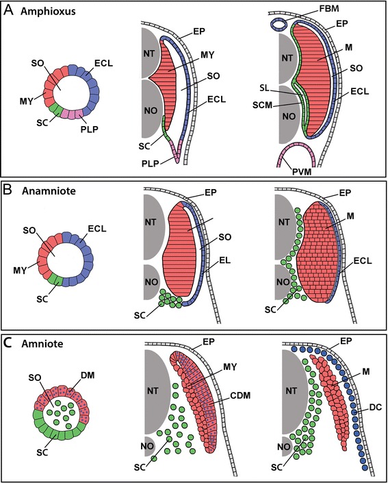
Figure 18.

Comparison of somite development and somite compartment derivatives in (A) amphioxus, (B) an anamniote vertebrate, and (C) an amniote vertebrate. For each, somite organization is schematized at early (left panels), mid (middle panels), and late (right panels) developmental stages. See text for details. The schematics are shown unbent from their true chevron- or W-shape and for simplicity omit the ribs and ventral muscles (and in anamniotes, the myoseptal cells), which are derived from the somites and migrate ventrally into the lateral plate mesoderm. Other details are also omitted, including the difference between epaxial and hypaxial musculature and between phasic and tonic muscle fibers. Abbreviations: CDM, central dermomyotome; DC, dermal cells; DM, dermomyotome; ECL, external cell layer; EP, epidermis; FBM, fin box mesothelium; MY, myotome; NT, neural tube; NO, notochord; PLP, presumptive lateral plate; PVM, perivisceral mesothelium; SL, sclerocoel; SCM, scleromesothelium; SO, somitocoel; SC, sclerotome; M, trunk muscles. Major synonyms that others have used for the foregoing structures are listed in Table 1.
