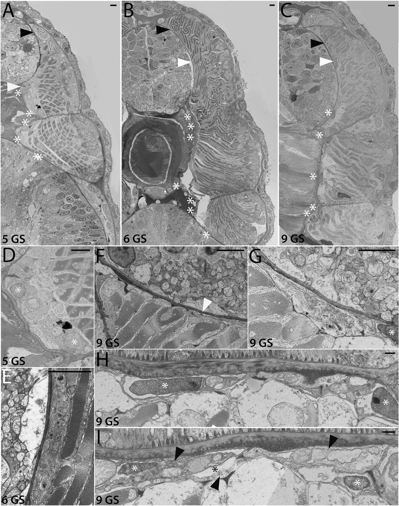
Figure 4.

Development of the non-myotome somite (2): mid to late larval stages. Transmission electron micrographs show the position of the myotome and the non-myotome (external cell layer and sclerotome) cells at the stages indicated. (A-C) are whole somite views that show the progressive change in position of the sclerotome. In these and panels, white arrowheads mark the border between sclerotome and myotome, and black arrowheads mark the border between the dorsal myotome and external cell layer. White asterisks show positions of sclerotome nuclei beside the midline structures. (D) is a detail of (A) showing the sclerotome-myotome boundary. (E) is a detail of (B) showing sclerotome beside the neural tube, (F-H) are details of (C) showing (F) the process of an external cell layer cell extended ventrally along the neural tube (arrowhead), (G) the nucleus of a sclerotome cell beside the notochord (H) a single-layered sclerotome along the notochord. (I) shows a different 9 GS specimen in which the sclerotome beside the notochord is double layered; black arrowheads in opposite directions indicate the two layers, black asterisk marks the sclerocoel. (A-E), dorsal is up, medial is to the left. (F-I) are rotated 90° counterclockwise, with dorsal to the left and medial up. Scale bars are 2 μm. Stage abbreviations: GS, gill slit.
