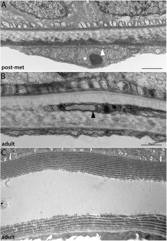
Figure 9.

Development of the dermis (3): postmetamorphic and adult stages. (A, B) Striated collagen layers continue to increase within the dermis during juvenile and adult stages. In all panels, epidermal ectoderm is up and mesothelium derived from the external cell layer is down. White arrowhead marks a clathrin-coated vesicle; white asterisk marks the nucleus of a mesothelial cell derived from the external cell layer. Fibroblast-like cells appear embedded within the dermal collagen during adult stages (black arrowhead), which, like the mesothelial cells below them, are rich in rough ER. (C) In subadults, a gelatinous layer is sometimes observed between the dermis collagen layers. When cut in the sagittal plane in (C), cross-hatching of the striated collagen is evident. A cutaneous canal within the gelatinous layer is visible on the left side of the panel. Scale bars are 1 μm. Stage abbreviations: post-met, post-metamorphic juvenile; adult, subadult.
