
Figure 3.
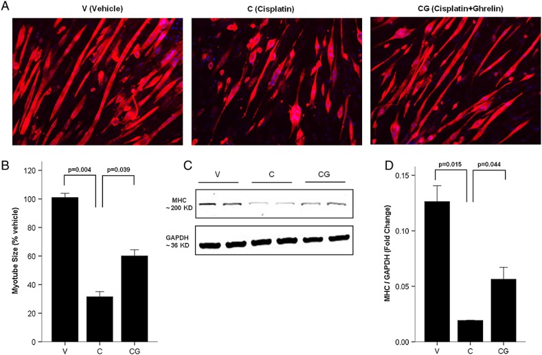
Ghrelin improves cisplatin-induced myotubes breakdown. (A) Immunofluorescence staining for anti-myosin/myosin heavy chain (MHC) antibody in C2C12 myotubes. MHC staining outlines the myotubes (red). 4'6-Diamidino-2-phenylindole was used to stain the nuclei (blue). (B) Myotubes size expressed as % from vehicle. (C–D) Western blot of MHC and glyceraldehyde-3-phosphate dehydrogenase (GAPDH) in C2C12 myotubes. V, vehicle-treated group; C, cisplatin-treated group; CG, cisplatin + ghrelin-treated group.