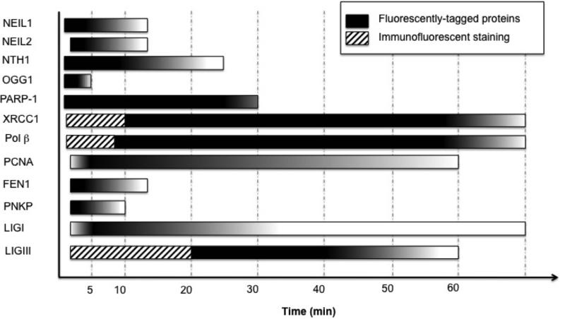
Figure 3. Coordination of BER proteins responding to sites of induced damage.
Time scales were estimated from all of the reports reviewed with peak recruitment shaded darkly and gradual dissociation of proteins illustrated by the color gradient. For PCNA and LIGI a slower recruitment was observed (19, 24), so a gradient reflecting this accumulation was included. The endpoints reflect the longest reported time monitored. Hashed blocks indicate recruitment detected by immunofluorescent staining.

