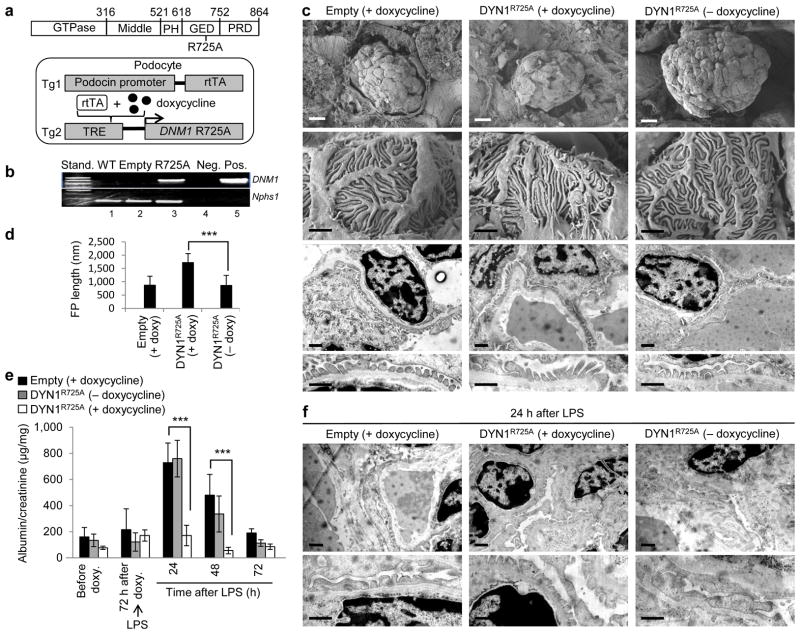
Figure 3.
Dynamin oligomerization in podocytes protects against proteinuria. (a) Domain structure of dynamin (top). R725A mutation is situated in the GED, which renders dynamin prone to oligomerize. A schematic diagram (bottom) indicating that the human gene DNM1 carrying R725A mutation (DNM1R725A) was placed under the regulation of a tetracycline-responsive promoter element (TRE; tetO). This transgenic mouse (Tg2) was subsequently bred to a second transgenic strain expressing the reverse tetracycline-transactivator (tTA) protein under the control of a podocin-specific promoter to allow for podocyte-specific gene expression (Tg1). Expression of DNM1R725A was induced by administration of the tetracycline analog, doxycycline. (b) RT-PCR of DNM1 from wild-type mice (WT), podocin-Cre only transgenic mice (Empty) fed with doxycycline and homozygous DNM1R725A/R725A transgenic mice (R725A) fed with doxycycline. Neg, negative control with water as a template; Pos, positive control with plasmid encoding DNM1R725A. Nephrin (Nphs1) was used as a positive control. (c) Representative electron micrographs of glomeruli (n = 5–6 glomeruli per genotype) from Empty and R725A transgenic mice fed with either a normal diet (− doxycycline) or doxycycline diet (+ doxycycline). Rows 1 (scale bars, 10 μm), and 2 (scale bars, 1 μm) show scanning electron microscopy. Rows 3 and 4 (scale bars, 1 μm) show transmission electron microscopy (TEM) images. (d) Length of foot processes determined by analyzing images in d. Doxycycline (doxy) (e) Proteinuria determined by analysis of spot urine samples at indicated times and in the indicated genotypes. Mice were fed with doxycycline (doxy) diet before they were injected with LPS (n = 6 mice per condition). Error bars, mean ± SD (***P ≤ 0.001, unpaired t-test). (f) Representative TEM images of glomeruli (n = 5–6 glomeruli per genotype) from Empty and R725A transgenic mice 24 hours after LPS injection. Animals were fed with either a normal diet (− doxycycline) or doxycycline diet (+ doxycycline). Scale bars, 2 μm (top row) and 1 μm (bottom row).

