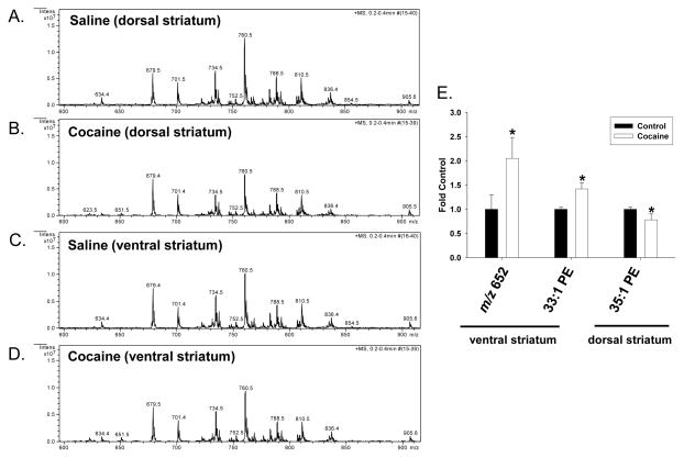
Figure 3. Effect of cocaine exposure on the relative abundance of phospholipids in the dorsal and ventral striatum.
Rats were treated with either saline or cocaine as described in Figure 1A. Seven days after the final treatment (Day 22) ventral and dorsal striatal tissue was isolated, subjected to Bligh-Dyer extraction and analyzed by ESI-MS. A. and C. represent positive ion ESI-MS spectra from control rats for the indicated tissues, while B. and D. represent spectra from cocaine exposed rats. E. represents changes in the relative abundance of select phospholipids in cocaine treated rats as compared to saline controls, and is presented as the mean ± the SD of at least 6 different rats. *Indicates a significant difference (p < 0.05) as compared to saline control rats. Lipid species indicated by only their m/z value were unable to be fully identified by subsequent MS/MS and neutral loss scanning.

