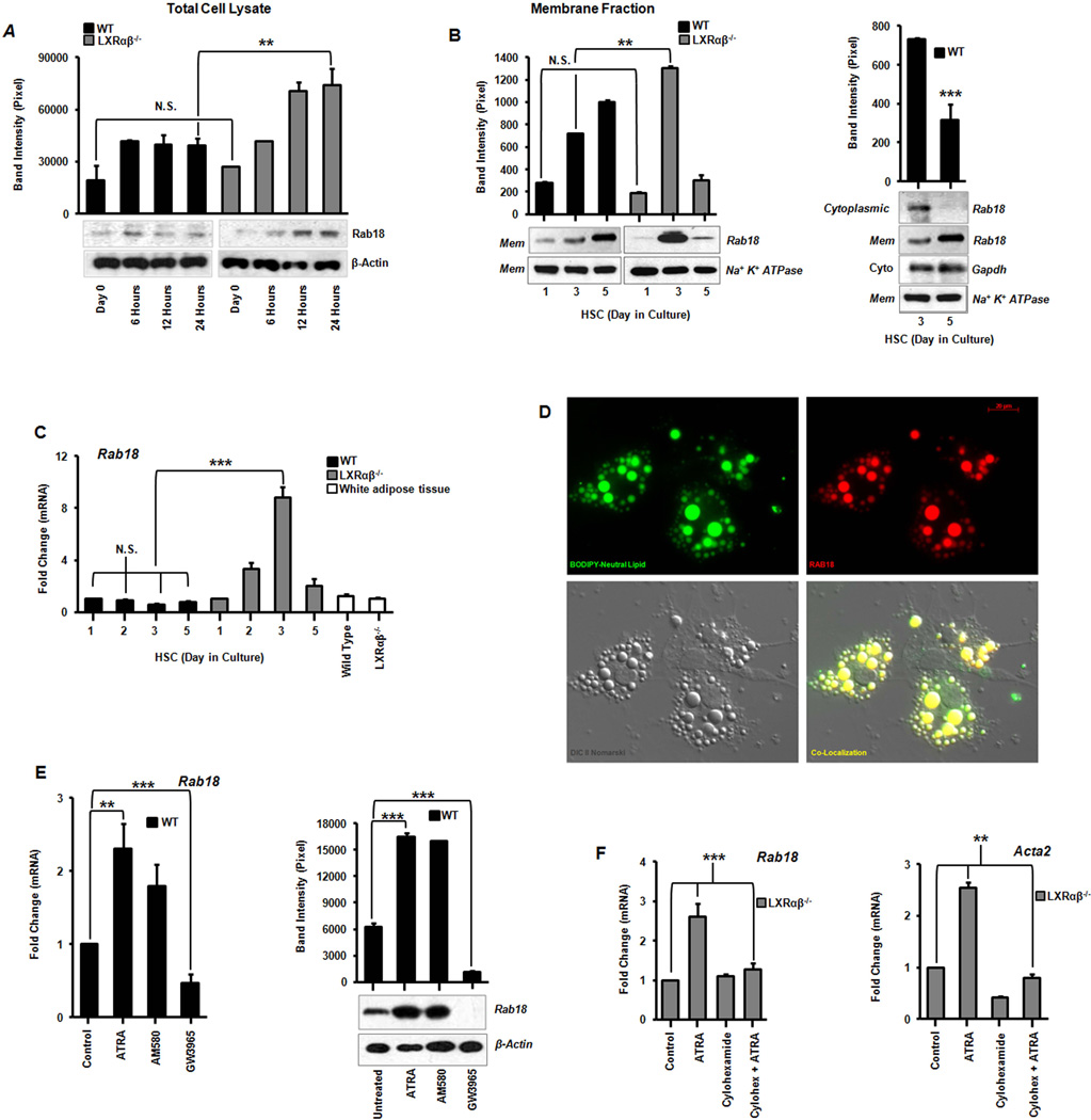
Figure 4. Identification of Rab18, a retinoid responsive lipid droplet associated protein.
(A) Rab18 protein expression by immunoblotting from total cell lysates and corresponding band densitometry in the first 24 hours of primary stellate cell culture activation. (B) Immunoblot analysis of Rab18 in HSC membrane and cytosolic fractions showing a shift from cytoplasm to membrane inserted Rab18 as activation proceeds in wild type stellate cells. (C) Rab18 gene expression in culture activated primary HSCs and compared to white adipose tissue (N=12 mice/genotype). (D) Immunofluorescence microscopy demonstrates Rab18 localization to LD surfaces. BODIPY (green), Rab18 (red), Nomarski DIC imaging, and the merged image bottom right; magnification 63×. (E) Rab18 mRNA and protein expression in day 2 culture activated primary WT HSCs treated with ATRA (100 nM), AM580 (100 nM), or GW3965 (1 µM). (F) Cycloheximide (10 µM) abrogates ATRA-induced Rab18 expression. All data are mean ± SEM, analyzed by 1-way ANOVA with post-hoc tests: *, P < .05; **, P < .01; ***, P < .001; NS, P > .05.

