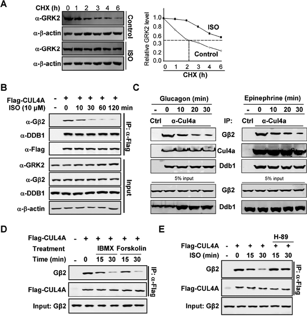
Figure 4. Activation of GPCR disrupts Gβ2 binding to DDB1.
(A) ISO stabilizes GRK2. HEK293 cells were treated with or without ISO, followed by CHX treatment as indicated time point. The protein levels of GRK2 were determined by Western blotting and quantified along with β-actin.
(B) Time dependent decrease of DDB1-CUL4A and Gβ2 binding by ISO. HEK293 cells were transfected with plasmids expressing Flag-CUL4A and then treated cells with ISO for indicated length of time. The levels of individual proteins and the protein-protein interactions were determined by Co-IP and Western analyses using indicated antibodies.
(C) Time dependent decrease of endogenous DDB1-CUL4A and Gβ2 binding by glucagon and epinephrine in cardiomyocytes.
(D) Dissociation of CUL4A and Gβ2 by IMBX/forskolin treatment. Flag-CUL4A was transfected into HEK293 cells and then treated the cells with IMBX/forskolin. The individual proteins were determined by Co-IP and Western blot analyses.
(E) PKA inhibitor H-89 blocks ISO effects on CUL4A-Gβ2 dissociation. Flag-CUL4A was transfected into HEK293 cells and then treated the cells with ISO/H-89. The individual proteins were determined by Co-IP and Western blot analyses.

