
Figure 2.
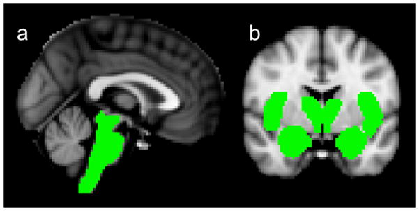
Selected vagal projections (“masked” in green) for the ROI analyses designed to test the hypothesis. a. Mask for the brainstem analysis. b. Masks for the forebrain analysis: thalamus, hypothalamus, amygdala, nucleus accumbens, insula (pictured in coronal slice), and (included but not pictured in coronal slice) paracentral lobule and hippocampus.