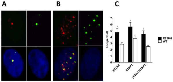
Figure 5. Expression of R280H hXRCC1 in MCF10A cells results in an increased level of double strand breaks.

Representative γH2AX/53BP1 immunostaining images of MCF10A expressing WT (A.) or R280H hXRCC1 (B.). Left upper panel represents γH2AX staining (red), right upper - 53BP1 staining (green), right bottom - DAPI staining (blue), and left bottom is a composite picture of all three panels (yellow for co-localizing foci). C. Only co-localizing foci (yellow on composite picture) were counted in WT or R280H hXRCC1 expressing cells. A total of at least 30 cells were scored for each cell line. Data are plotted as mean ± S.E. ** denotes p < 0.01.
