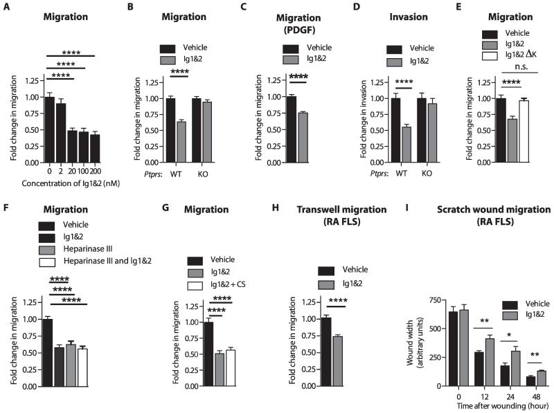
Fig. 2. RPTPσ Ig1&2 inhibits FLS migration and invasiveness in an RPTPσ-dependent manner.
(A) FLS migrated through Transwells in response to 5% FBS in the presence of the indicated concentrations of Ig1&2. (B and C) Migration of WT (B and C) or Ptprs KO FLS (B) through Transwells in response to (B) 5% FBS or (C) PDGF-BB (50 ng/ml) in the presence of 20 nM Ig1&2 or vehicle. (D) Invasion of WT or Ptprs KO FLS through Matrigel in response to 5% FBS in the presence of 20 nM Ig1&2 or vehicle. (E) Migration of FLS through Transwells in response to 5% FBS in the presence of vehicle or 20 nM Ig1&2 or non-GAG binding Ig1&2 ΔK mutant. n.s., not significant. (F and G) FLS migrated through Transwells in response to 5% FBS in the presence of (F) vehicle or 20 nM Ig1&2, with or without 10 mU of heparinase III pretreatment, or (G) vehicle, 20 nM Ig1&2, CS (100 μg/ml), or both Ig1&2 and CS. (H) RA FLS migrated through Transwells in response to 5% FBS in the presence of vehicle or 20 nM Ig1&2. (I) RA FLS monolayers were serum-starved before scratch-wounding and stimulated with 10% FBS in the presence of 20 nM Ig1&2 or vehicle. Wound width was measured at three points at the indicated times. (A to H) Mean ± SEM fold change of invasion or migration relative to vehicle-treated cells. Data are from three independent experiments (n = 45 fields; ****P < 0.0001, Mann-Whitney). (I) Mean ± SEM wound width from two independent experiments (n = 6 fields; 12 hours, **P = 0.0087; 24 hours, *P = 0.0411; 48 hours, **P = 0.0043; Mann-Whitney).

