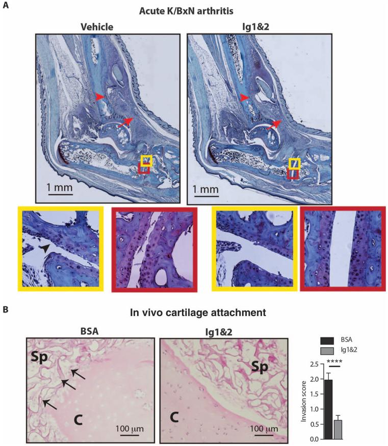
Fig. 7. RPTPσ Ig1&2 decreases FLS attachment to cartilage in vivo.
(A) BALB/c mice were induced with acute K/BxN arthritis and treated with vehicle or 0.5 mg of Ig1&2 intravenously daily (days 0 to 6). Pathology of safranin O–stained ankle sections from mice on day 14 of arthritis. Arrow indicates inflammation; arrowhead indicates bone erosion. FLS crawling over cartilage (arrow) (yellow insets) and reduced chondrocyte layer (red insets) in (left) vehicle-treated versus (right) Ig1&2-treated mice. Cartilage PG content (red insets) assessed by safranin O staining. (B) SCID mice were implanted subcutaneously with cartilage explants and RA FLS in surgical sponge and treated with BSA or Ig1&2 intravenously for 35 days. (Left) Representative images showing cartilage (C) and sponge (Sp). Arrows indicate FLS attaching to and invading cartilage. (Right) Mean ± SEM invasion scores; n = 3 mice per group (n = 30 fields; ****P < 0.0001, Mann-Whitney).

