Figure 6.
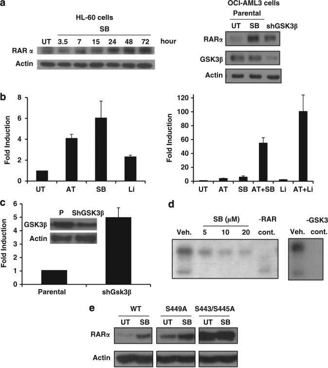
Mechanisms through which GSK3 modulates RAR signaling. (a) GSK3 inactivaton induces RARα expression. First panel: HL-60 cells were treated for the indicated times with SB (30 μm) and western analysis was performed. Second panel: OCI-AML3 cells in which GSK3β was stably knocked down demonstrate an increase in RARα expression as compared with empty vector infected parental cells. The parental cells also exhibit an induction in RARα expression after 72 h of SB (30 μm) treatment. (b) GSK3 inhibition enhances RAR transcriptional activity. Hela cells transfected with RARE-luc (RAR response element-luciferase) were treated for 16 h with ATRA (1 μm), SB (30 μm), lithium (20 mm) or a combination and the luciferase signal was measured. The single treatments are shown in a separate graph to demonstrate the induction of RARE with GSK3 inhibition alone. (c) GSK3β knockdown induces RAR transcriptional activity. Parental and GSK3β knockdown Hela cells were transfected with RARE-luc and assessed for luciferase activity after 72 h. (d) GSK3β phosphorylates RARα. Recombinant GSK3β phosphorylates recombinant RARα (as well as itself) using a P32 labelled in vitro kinase assay. GSK3 autophosphorylation and RAR phosphorylation are inhibited by SB treatment. (e) Mutant RAR shows increased expression and a loss of GSK3-mediated RAR induction. Hela cells were transfected with wild-type RAR, a S449A RAR mutant or a S443A/S445A RAR mutant and assessed for RAR protein expression with and without SB treatment (30 μm) for 24 h. Transfection efficiency was found to be equal with these constructs. The cells were all co-transfected with prL-CMV (Promega) and the Renilla luciferase signal was measured to control for transfection efficiency (data not shown).

