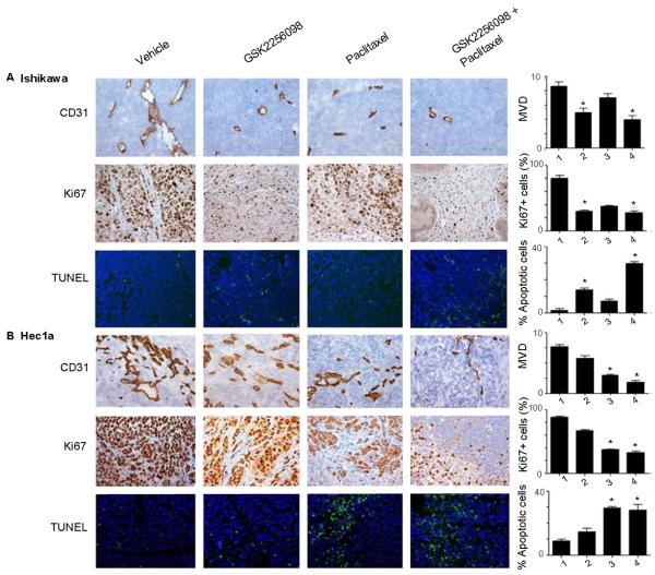
Figure 4. Effect of GSK2256098 in vivo on angiogenesis, proliferation, and apoptosis in uterine tumors.
(A) Ishikawa and (B) Hec1A tumors collected at the conclusion of in vivo therapeutic experiments were subjected to CD31, Ki67, and TUNEL staining. Representative sections (final magnification, ×200) are shown for the four treatment groups (vehicle control, GSK2256098, paclitaxel, and a combination of GSK2256098 and paclitaxel). The average number of CD31-positive vessels per field, mean percentage of Ki67-positive cells (proliferative index), and mean percentage of TUNEL fluorescence-positive cells (percentage of apoptotic cells) are shown in the adjoining graphs. Five fields per slide and at least five slides per treatment group were examined and compared using the Student t-test and analysis of variance. *P < 0.05 versus control.

