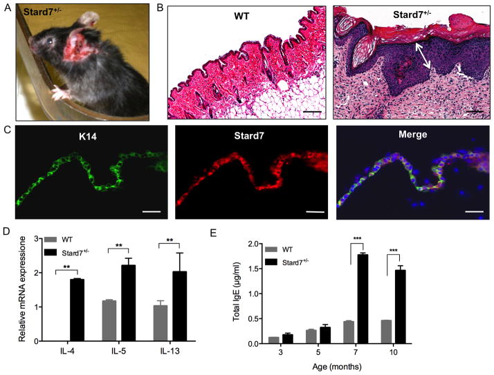
Fig. 7. Stard7+/− mice develop spontaneous dermatitis.
(A) A skin lesion on the ear of a 7 month-old Stard7+/− mouse. (B) H&E staining of skin tissue from 7 month-old WT and Stard7+/− mice. A section through the skin lesion shows expansion of basal, spinous, granular and cornified layers of the epidermis (white arrow) and cellular infiltration of the dermis in Stard7+/− mice. Scale bars = 50 μm. (C) Immunofluorescence of the epithelial cell marker K14 and Stard7 in skin tissue from 8 week-old WT mice. Scale bars = 25 μm. (D) qRT-PCR of Th2 cytokine mRNAs in skin tissue of 7 month-old WT and Stard7+/− littermates. β-actin was used as an endogenous control. n = 3 mice/group. (E) Total IgE in the serum of mice over time. n = 4 mice/group.

