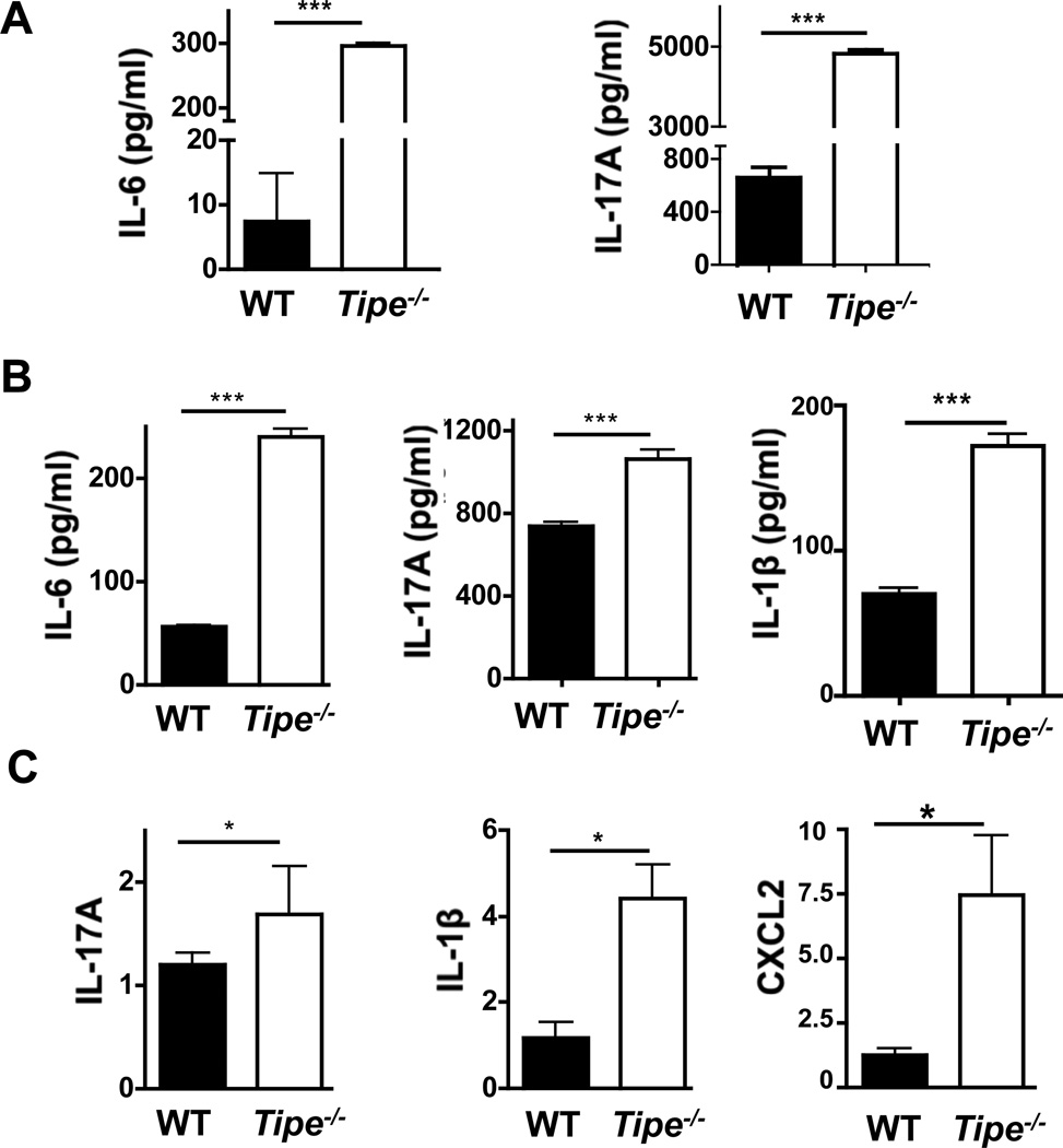
Figure 4. Increased inflammatory cytokines in serum and colon of Tipe−/− mice.
WT and Tipe−/− (n=5) mice were treated with DSS water for five days, followed by regular drinking water for two days. Mice were then sacrificed, and their blood was collected retro-orbitually. Colons were harvested and ½ of them were homogenized in lysis buffer (weight/volume = 1/10). The cytokines in sera and colon homogenates were measured by ELISA. (A) IL-6 and IL-17 -in sera. (B) IL-6, IL-17, and IL-1β in colon homogenates. (C) Total RNA was isolated from the other half of the colon using Trizol reagent. The levels of IL-17, IL-1β, and CXCL2 mRNAs were examined using quantitative real-time PCR. Data shown are representatives of two experiments. *P<0.05; **P<0.01; *** P<0.001.

