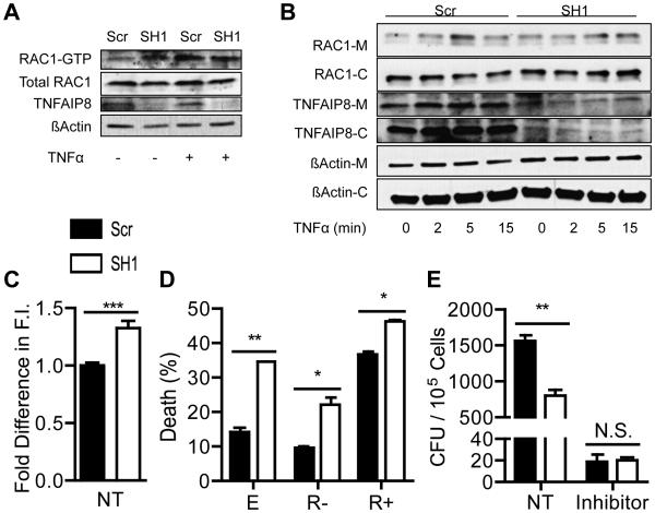
Figure 4. TNFAIP8 and RAC1 in L. monocytogenes infection and cell death.
The levels of RAC1-GTP was determined by a PAK-GST pull down assay for control and TNFAIP8 knockdown Hepa1-6 cells stimulated with or without 50 ng/ml of TNFα for 5 minutes. Relative band intensities: 1, 1.5, 1.7, and 1.54 for Scr and SH1 with no treatment, and Scr and SH1 with TNFα treatment, respectively. (B) Control and TNFAIP8 knockdown Hepa1-6 cells were stimulated with 50 ng/ml of TNFα for 0, 2, 5, or 15 minutes. Lysates were fractionated into membrane [M] and cytoplasmic [C] portions, and TNFAIP8, RAC1, and actin were detected by Western blot. Relative band intensities: 1, 1.61, 2.86, 1.84, 1.72, 1.42, 1.91, and 2.04 for Scr with 0, 2, 5, and 15 minutes of TNFα treatment and SH1 with 0, 2, 5, and 15 minutes of TNFα treatment, respectively. Band intensities were determined by ImageJ software. (C) Control and TNFAIP8 knockdown Hepa1-6 cells were stained with phalloidin-FITC to quantify their total levels of F-actin by flow cytometry. (D) Control and TNFAIP8 knockdown Hepa1-6 cells were transiently transfected with pEGFP vectors containing either the EGFP cDNA alone [E], or EGFP plus RAC1-17N [R-] or RAC1-61L [R+] cDNAs. Cells were treated with 5 ng/ml of TNFα and 20 ng/ml of cycloheximide for 6 hours. EGFP positive cells were gated by flow cytometry and cell death was detected by Annexin V and 7-AAD staining. (E) Control and TNFAIP8 knowdown Hepa1-6 cells were infected with 50 MOI of L. monocytogenes for 1 hour with or without RAC1 inhibitor II [RI] Z62954982 at 100 μM. Cells were washed and then treated with 150 μg/ml Gentamycin for 30 minutes to kill extracellular bacteria before being lysed to release the intracellular bacteria. The results are representative of at least two independent experiments. * P < 0.05; ** P < 0.01; *** P < 0.001.

