
Figure 4. Gut-associated lymphoid tissue (GALT) lymphoid micro architecture in NSG and NSG-A2 mice reconstituted with umbilical cord blood or fetal liver hematopoietic stem cells.
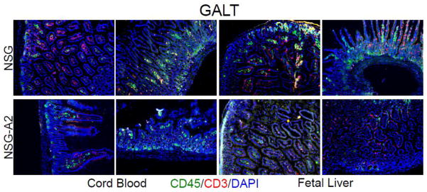
Intestines were isolated from either umbilical cord blood (left panels) or fetal liver (right panels) stem cell reconstituted mice of the indicated types, processed for histologic analysis, and sections stained with fluorescence antibodies specific for the indicated markers as well as DAPI to delineate all cell nuclei. Stained sections were analyzed by fluorescence microscopy. Note the rather low human immune cell reconstitution in the lamina propria and villi regions in umbilical cord blood or fetal liver stem cell reconstituted NSG-A2 intestines (lower panels) as compared to the well reconstituted areas in the intestines of NSG mice. Data are representative of those obtained from at least two reconstituted mice of each type.