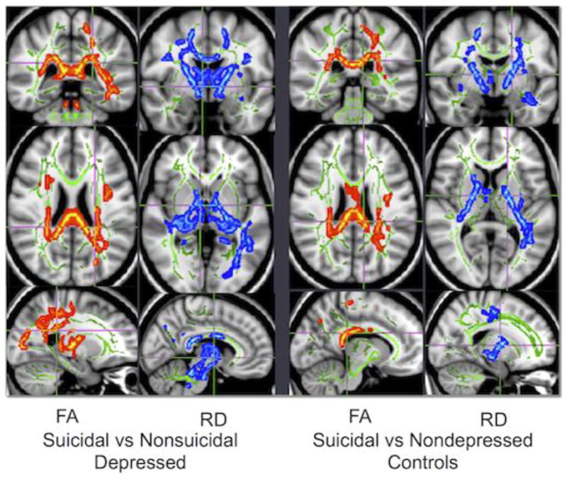
Figure 3. Voxelwise comparison of DTI measures between groups.

Comparisons between groups in whole-brain measures of FA (fractional anisotropy) and RD (radial diffusivity). Please refer to Table 2 for location of cluster maxima. Green voxels represent the white matter skeleton, red voxels are areas of lesser value in the suicidal group, and blue voxels are areas of greater value in the suicidal group. In these analyses, the suicidal group exhibited lower FA and higher RD in identified regions when compared with either the nonsuicidal depressed group or the control group. There were no white matter regions exhibiting significant differences in either FA or RD between the nonsuicidal depressed and nondepressed control groups.
