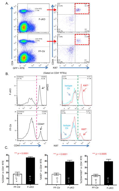
Figure 1. Newly released CD4+ T cells from the atrophied thymus acquired an activated immune cell phenotype.
F-cKO and FF-Ctr (fx/fx-uCreERT and fx/fx mice, respectively) were crossed with Rag2-GFP mice and then FoxN1fx/fx deletion was induced in 6-week-old adult mice with i.p. injected tamoxifen (TM). 14 days later, peripheral splenocytes were freshly isolated and stained with CD4, CD44, and Ki67 antibodies, and CD4+GFP+ cells were defined as CD4+ RTEs. (A) Representative dot plots show CD44hiKi67+ cell gates (red boxes) in CD4+ RTEs from F-cKO (top panels) and FF-Ctr control (bottom panels) mice. (B) Representative histograms of CD44hi (left panels) and Ki67+ (right panels) in CD4+ RTEs from F-cKO (top panels) and FF-Ctr control (bottom panels) mice. (C) Summarized results of % CD44hi, Ki67+, and CD44hiKi67+ double positive cells in CD4+ RTEs (from left to right panels). A Student t-test was used to determine statistical significance between groups. All data are expressed as mean ± SEM. Data are pooled from at least three independent experiments (n = animal numbers).

