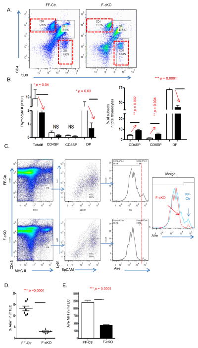
Figure 3. Clonal deletion of SP thymocytes was impaired in the FoxN1-cKO atrophied thymus.
(A and B) Five days after inducing FoxN1fx/fx deletion with TM in 6-week-old FF-Ctr and F-cKO mice, thymoyctes were freshly isolated for cell surface staining of CD4 vs. CD8. (A) Representative flow cytometry plots show CD4+ and CD8+ SP gates of F-cKO (right panel) and FF-Ctr control (left panel) mice. (B) Summarized results of absolute cell numbers per thymus (left panel) and % of each subpopulation in total thymocytes (right panel). Open bars represent FF-Ctr control mice, filled bars represent F-cKO mice. Each group contained seven animals. (C and D) Thymic epithelial cells were enzymatically digested from F-cKO and FF-Ctr thymi and intracellularly stained for Aire antibody. (C) Flow cytometry was used to gate on mTECs (CD45−, MHC-II+, EpCAM+, Ly51−), (D) Summarized results of the percentage of Aire+ mTECs, and (E) the mean fluorescent intensity of Aire+ mTECs. A Student t-test was used to determine statistical significance between groups. All data are expressed as mean ± SEM. Data are pooled from at least three independent experiments with a total of n = 7 animals per group.

