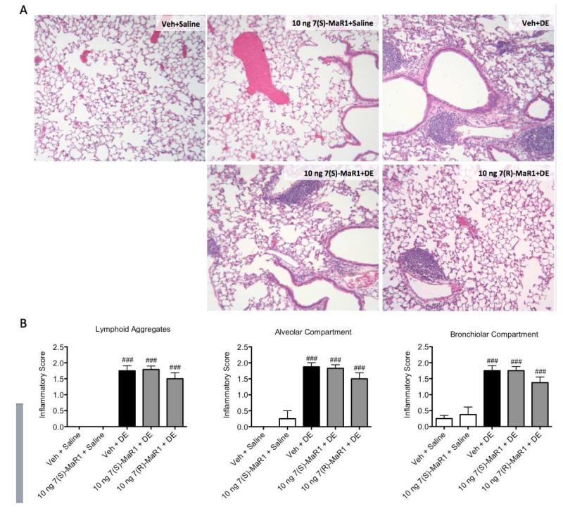
Figure 7. Lung histopathology following repetitive DE exposures in mice receiving pretreatment with MaR1.
Mice were pretreated with 0 or 10 ng MaR1 given i.p. 30 minutes prior to an intranasal dose of DE, given daily for 15 consecutive weekdays. At five hours following the final MaR1/DE exposure, lungs were formalin-fixed and inflated for paraffin embedding and sectioning. Lungs were stained with hematoxylin and eosin (A), then scored by a lung pathologist for markers of chronic inflammation (B). ### p < 0.001 compared to vehicle + saline group. Data are represented as mean values with standard error bars. N ≥ 4 mouse lungs used in analyses for control groups and N ≥ 8 mice for treatment groups.

