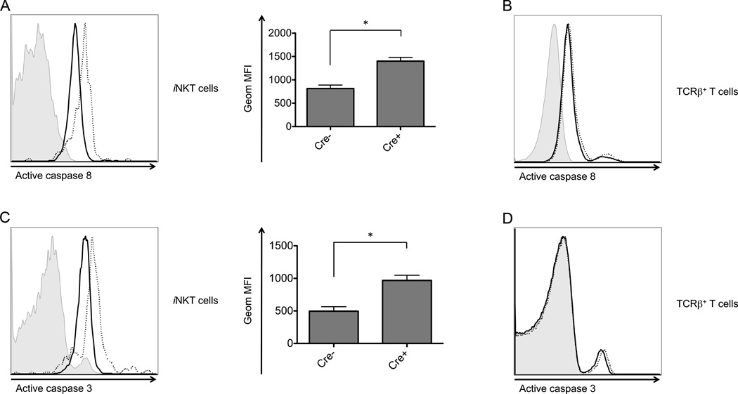
Figure 7.
Atg5 deficient thymic iNKT cells exhibited an increase in active caspase 8 and caspase 3. Atg5 deleted thymocytes were cultured overnight, followed by analysis by flow cytometry of active caspase 8 (A and B) and active caspase 3 (C and D) in thymic iNKT cells (A and C) and TCRβ+ thymocytes (B and D). Shown here are both flow cytometry plots (left) and geometric MFI (right). Data are representative of at least two independent experiments. Filled histogram: isotype control; solid line: wild type cells; dotted line: Atg5 deficient. *p<0.05, n≥3. Error bars are SD.

