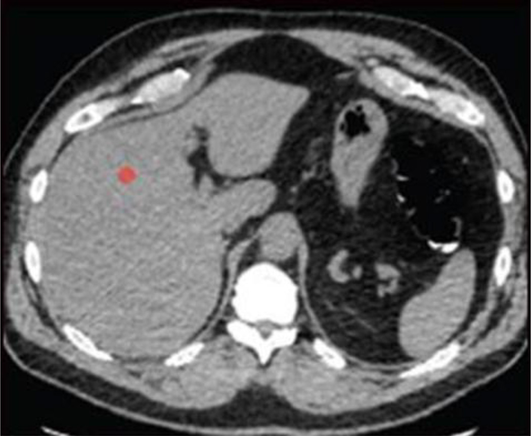
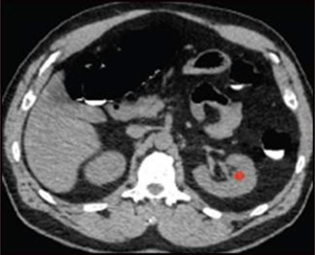
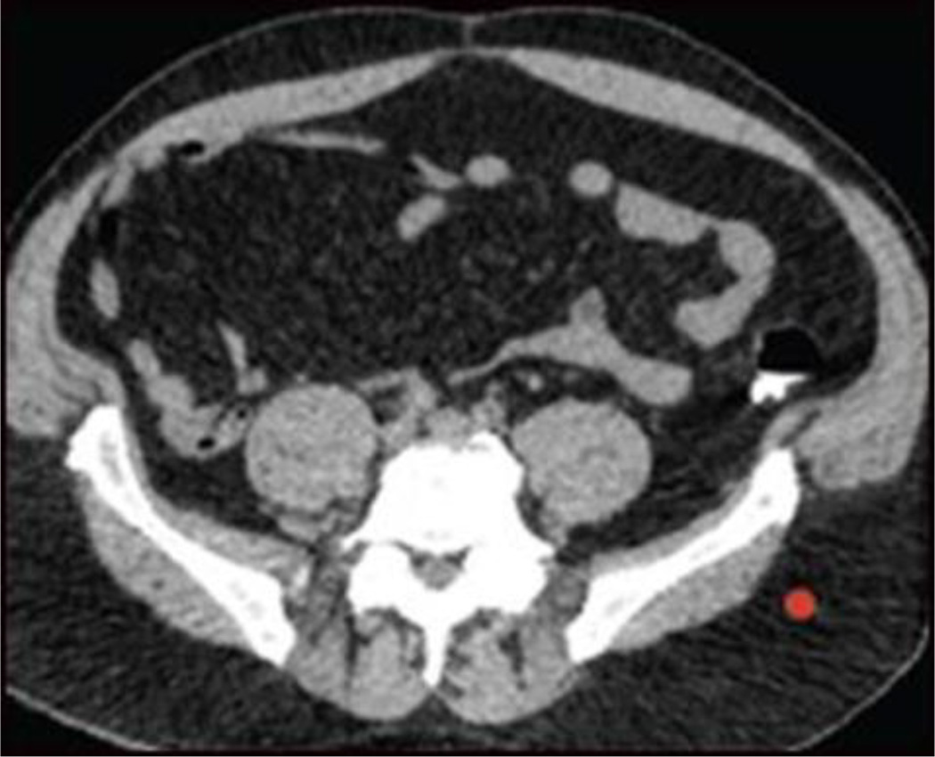
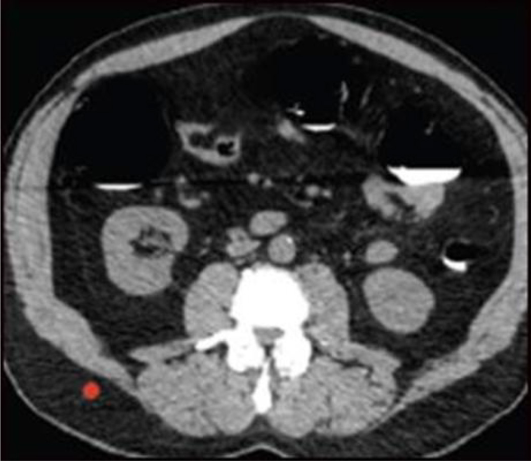
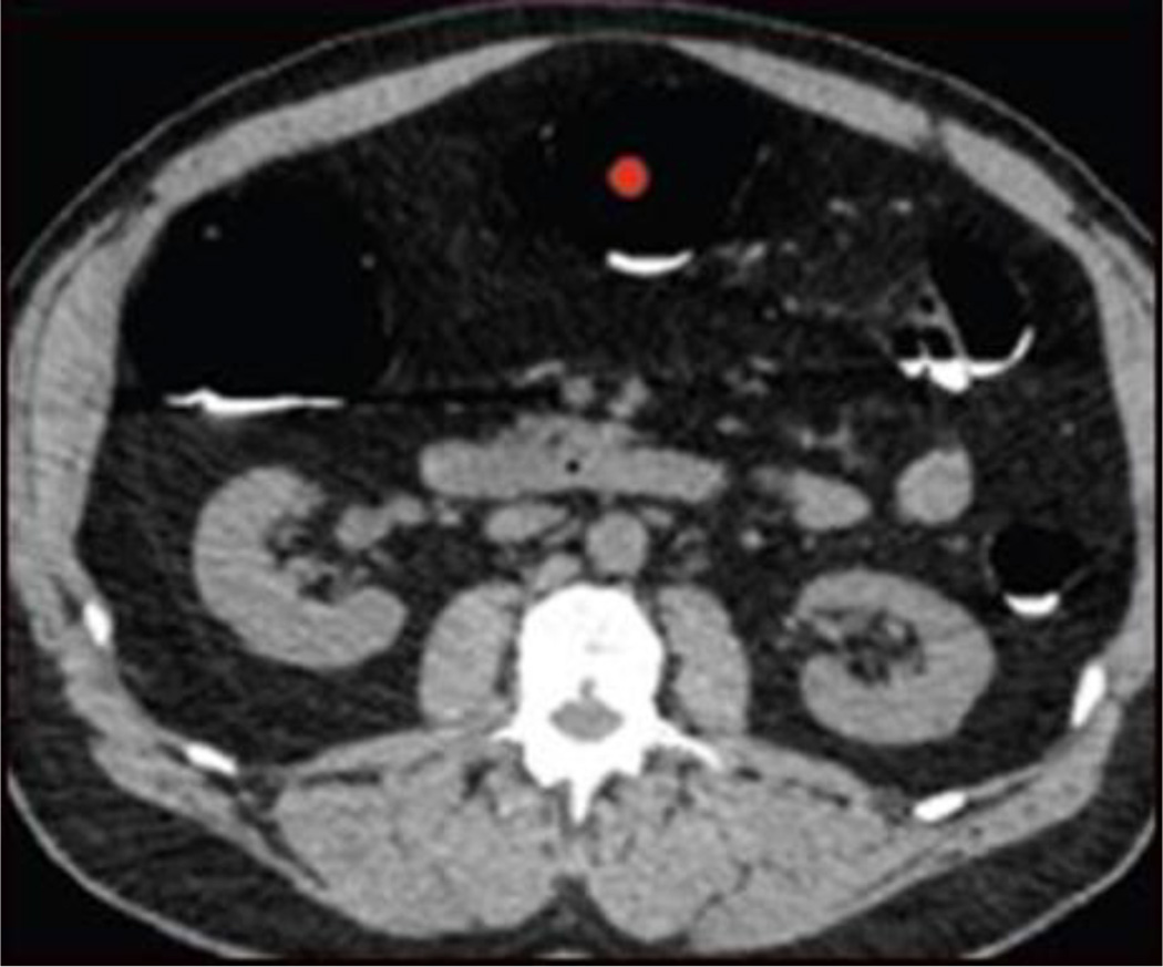
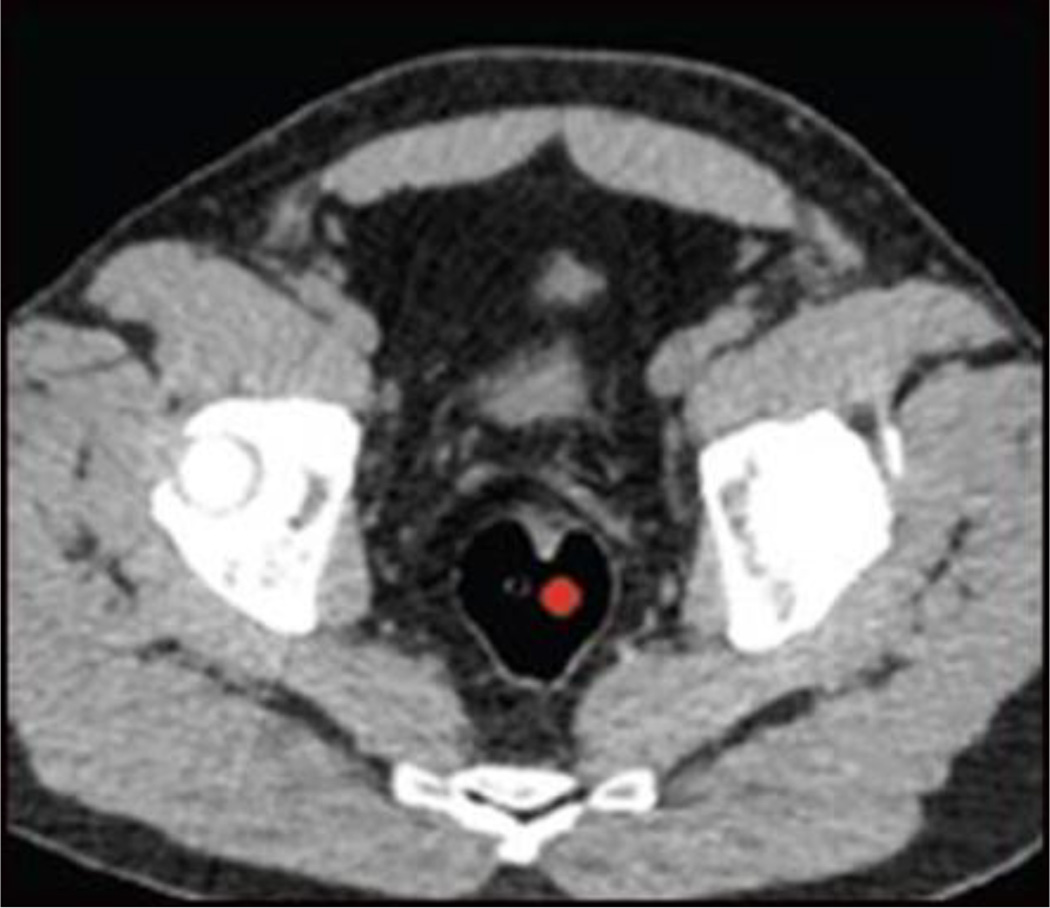
Figure 1.
ROI placement for objective noise measurement. Small red circles indicate location of ROIs, with two parenchymal sites (liver, A; left kidney, B), two fat attenuation sites (subcutaneous fat of left flank, C; right flank, D), and two colonic air column sites (transverse colon, E; rectum, F). ROIs were placed in homogeneous areas without vessels, soft tissue stranding etc.

