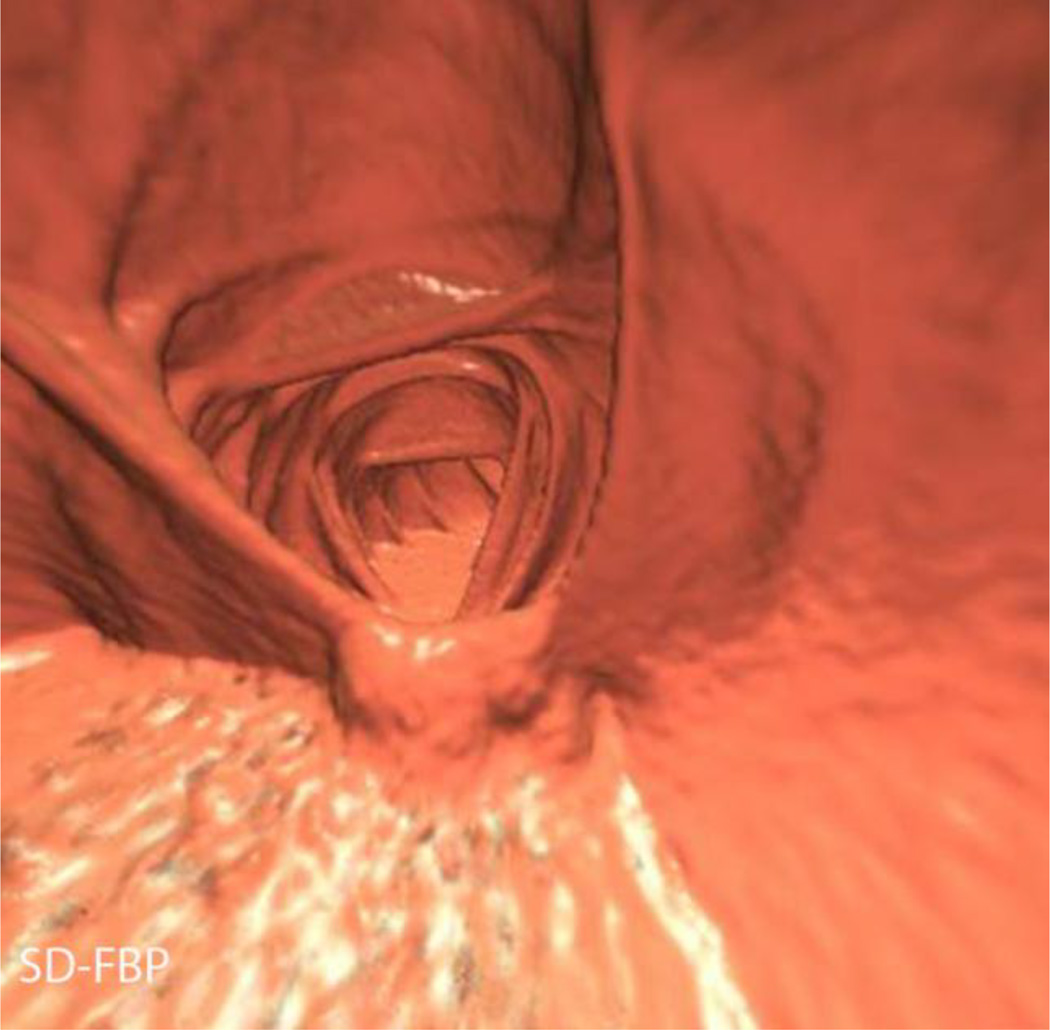
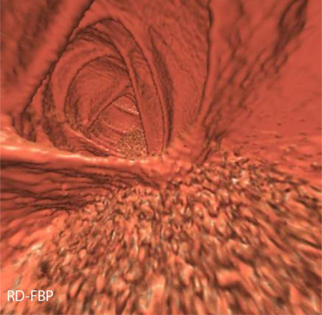
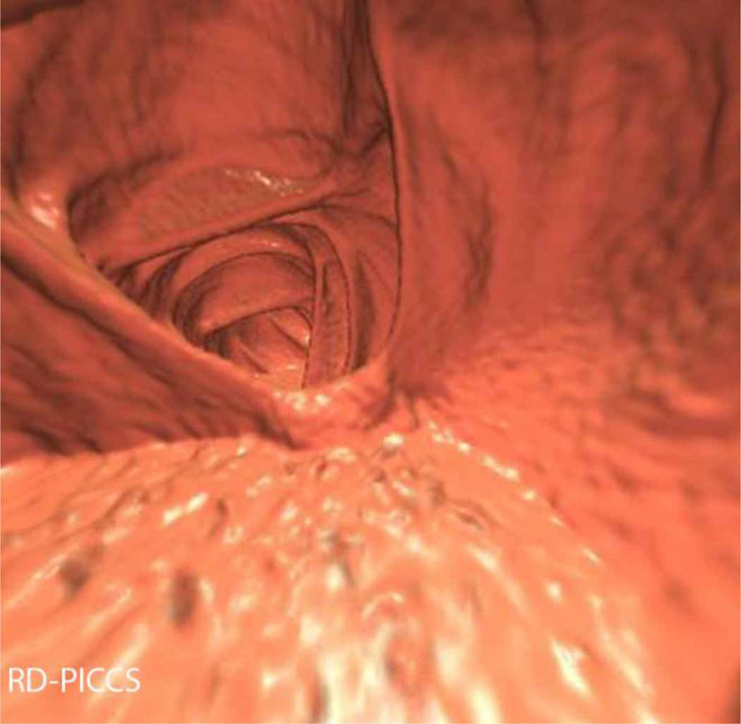
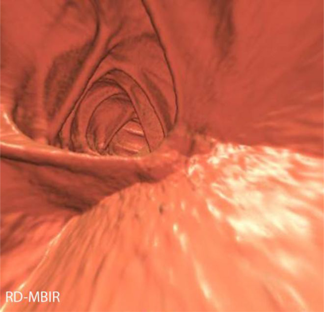
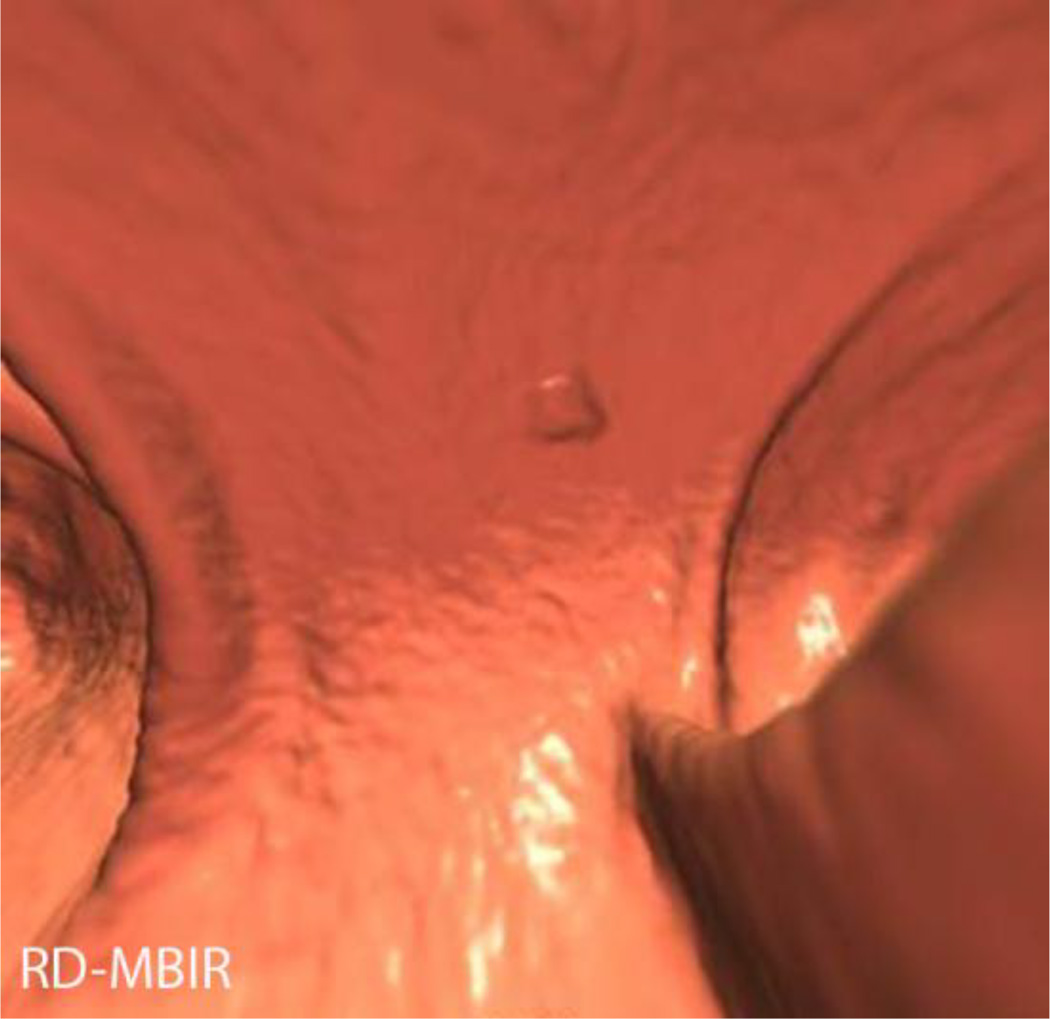
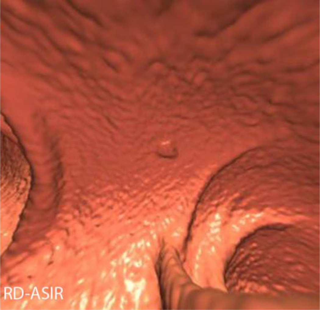
Figure 7.
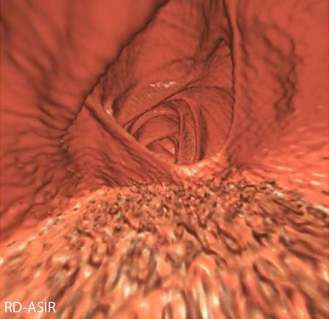
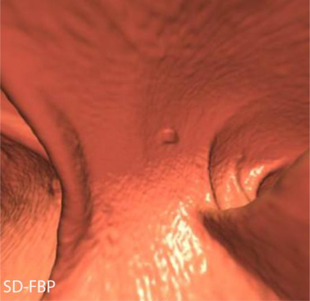
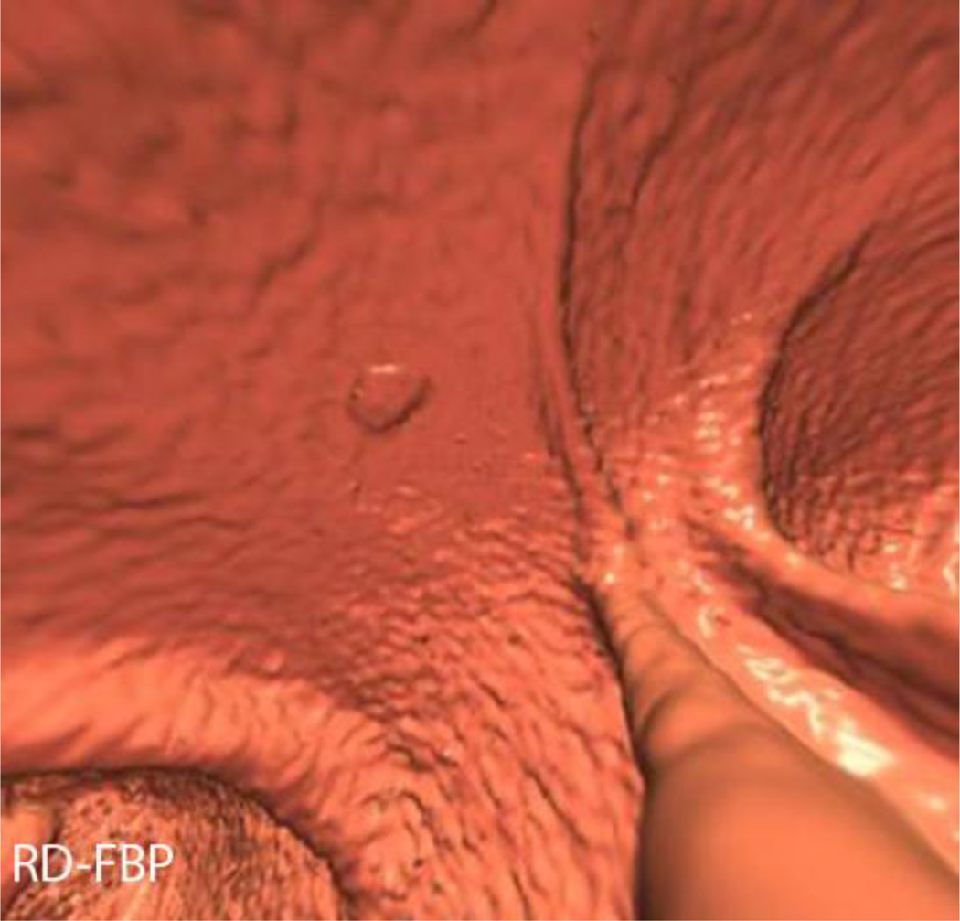
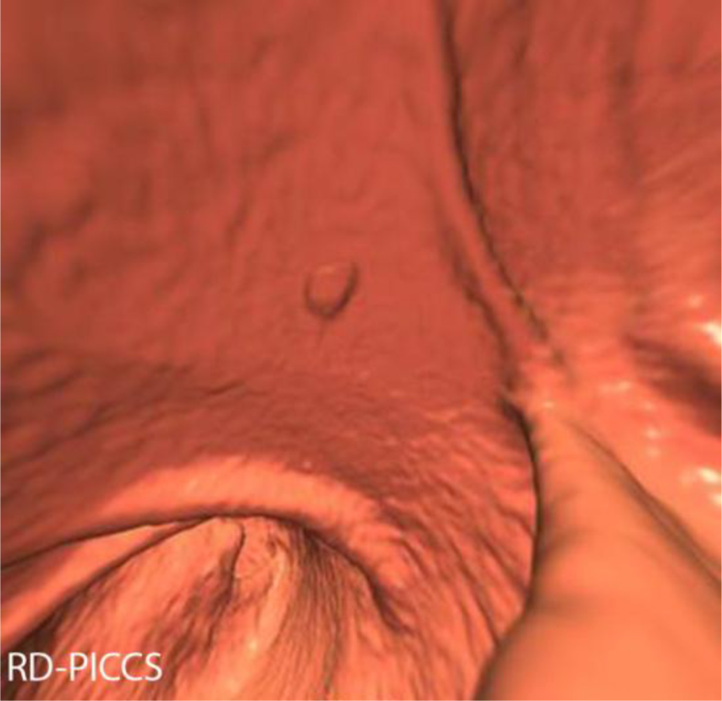
3D image quality assessment at the ileocecal valve. 3D images from CTC in a 53 year old male with BMI of 31.3, effective dose (SD) of 2.33 mSv, effective dose (RD) 0.48 mSv. Like with the 2D image quality scoring, the standard dose images (A) received the highest image quality scores, followed by RD-MBIR (D), RD-PICCS (C), which were scored significantly higher than the RD-ASIR (E) and RD-FBP (B). This same patient also had a 7 mm sigmoid tubular adenoma, shown here on SD-FBP (F), RD-FBP (G), RD-PICCS (H), RD-MBIR (I) and RD-ASIR (J). The SD-FBP (F), RD-PICCS (H) and RD-MBIR (I) received similar polyp conspicuity scores, higher than RD-FBP (G) and RD-ASIR (J).

