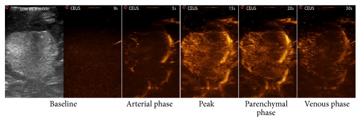
Figure 7.

CEUS phases. In this picture, a low mechanical index (MI) B-mode scan is depicted together with screenshot of the main phases of contrast enhancement dynamics. In the arterial phase, the main feeders are clearly visible. In peak and parenchymal phase, it is possible to differentiate hyper- or hypovascularized areas within the tumor. In the venous phase, multiple small draining vessels are recognizable.
