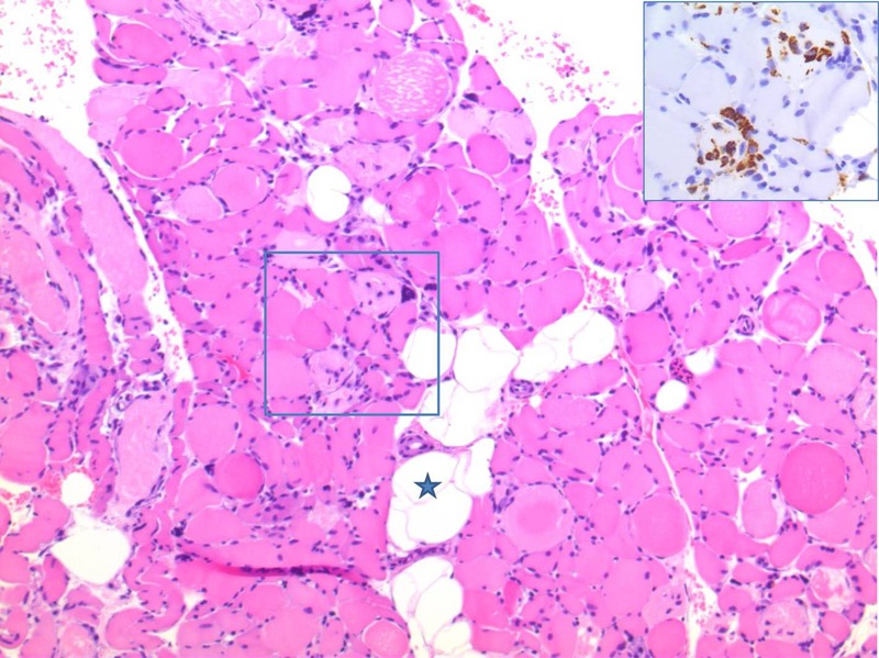
Figure 1.

Muscle biopsy with fatty infiltration (asterisk). There is variation in muscle fibre size, and many fibres are necrotic. Some of the degenerating fibres are undergoing myophagocytosis with infiltrating macrophages (H&E staining). Insert: Parallel section stained with CD68, highlighting the infiltrating macrophages.
