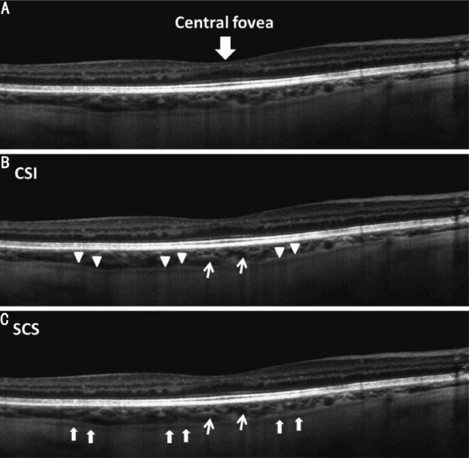
Figure 1. A single 9 mm horizontal line scan with enhanced depth imaging obtained from the right eye of a 55-year-old healthy male.
A: The choroidal-scleral interface (CSI); B: Is indicated by a well demarcated hyper-reflective band (triangle heads) and the suprachoroidal space (SCS); C: Is indicated by the hypo-reflective band (arrows). The narrow arrows indicate the cross section of large choroidal vessels.

