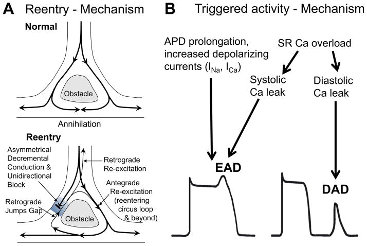
Figure 2.
Proarrhythmogenic mechanisms. A) Reentry. Upper panel shows normal conduction around an obstacle. The latter exhibits electrical properties that differ dramatically from the rest of the myocardium (slowed conduction and/or repolarization). The wavefront propagates around this line of block and reaches the back from multiple directions simultaneously resulting in wave annihilation. If, however, unidirectional block (often coupled with decremental conduction) exists around one side (lower panel), the wavefront can reenter and re-excite tissue in front of the unidirectional block. This can lead to stable reentry. B) Triggered activity. Increased depolarizing currents that result in prolongation of action potential duration (APD) can favour L-type Ca channel reactivation. Increased Ca entry via ICa can also lead to SR Ca overload and spontaneous SR Ca release during AP plateau phase. This may generate transient inward (ITi) current by Na/Ca exchange. Both Ca channel reactivation and ITi may depolarize the membrane during AP plateau phase, which can result in an early afterdepolarization (EAD). In addition, SR Ca overload may also result in diastolic SR Ca release. The consequent ITi may lead to depolarization from the resting membrane potential, which can result in a delayed afterdepolarization (DAD). Partially reproduced.31

