
Fig. 2.
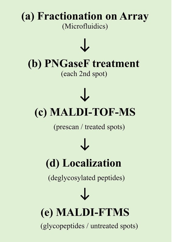
Workflow for the site-specific glycosylation analysis as used for IgM (a-e). After fractionation of the eluent from the nano-LC separation (a), a specific portion of the collected fractions (every other spot) was digested using PNGaseF (b). Subsequently, a first low-resolution MALDI-TOF-MS scan in the lower mass range of the PNGaseF-treated spots was performed (c). After the localization of the deglycosylated peptides in the initial data set (d), a high-resolution MALDI-FTMS scan (at a high mass range) was performed solely on the selected untreated spots.