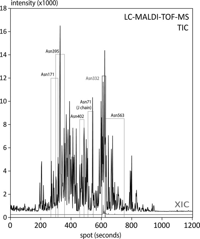
Fig. 3.

Upper trace: TIC of the treated spots (each 2nd spot on the chip) generated by the lower-resolution scan using a MALDI-TOF-MS instrument. The elution areas of the deglycosylated peptides from individual sites are indicated with boxes, which were identified by the appearance of the deglycosylated peptide after PNGaseF treatment. The glycopeptide for Asn 563 provided a broad tailing peak (broad box). Lower trace (red): Example for an extracted ion chromatogram (XIC) from the treated spots, as shown for the deglycosylated peptide from glycosite Asn 332 ([M+H+]+ = 2340 Da).
