. 2015 May 13;5:tre-5-301. doi: 10.7916/D8T72G9G

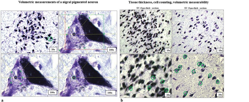
Figure 1A/1B. Quantification of SN Pigmented Neurons. The quadrants in the figure show how a randomly sampled pigmented neuron (first upper-right quadrant, 20× objective), and 6 randomly projected rays on that pigmented neuron intersect its cell body, nucleus, and nucleolus (respectively, upper-right, lower-left, and lower-right quadrant). A 100× oil immersion objective was used for all volumetric measurements and for each pigmented neuron. The green markers in each quadrant indicate the point of measurement from which the areas of the cell body, nucleus, and nucleolus were calculated. The colored square upper-right quadrant represents the “counting frame” used for the Optical Fractionator probe. The Stereo-investigator software automatically calculates the volumes of each single neuron computing various parameters such as the thickness of the tissue, the disector height, and guard zones. Figure 1B. Tissue thickness, cell counting, volumetric measurability of nigral pigmented neurons. SNc from a C brain (case#16), cut at different levels of thicknesses (40 µm and 10 µm), stained with CV, and inspected at two different magnifications (10× and 20× objectives). The green circles in the inferior parts of the figure indicate the number of pigmented neurons measurable when pigmented neurons were sampled using 40 µm-thick and 10 µm-thick tissue sections, respectively, with a 20× objective. Notably, the number of measurable pigmented neurons in SN cut at 10 µm of thickness (right of figure) is markedly higher due to the increased number of clear visible CV-stained nucleoli. The nucleolus was the established point of spatial reference on this study. All measurements were performed using a 100× oil-immersion, NA 1.30, neofluor ∞/0.17 objective. Abbreviations: C, Control; CV, Cresyl Violet; SN, Substantia Nigra; SNc, Pars Compacta of Substantia Nigra.

Figure 1A/1B