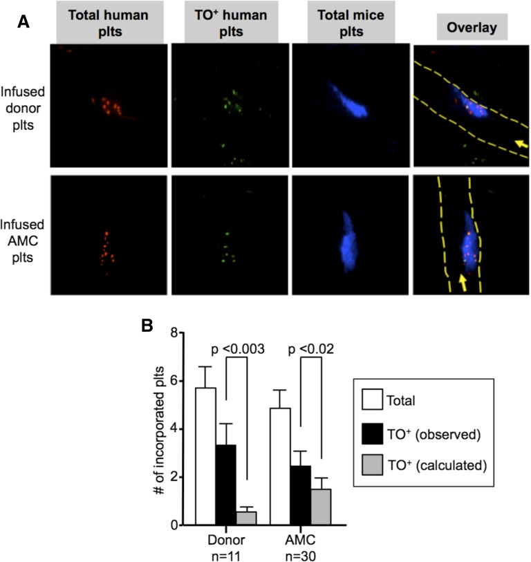
Figure 6.
In situ studies of incorporation of TO+ vs TO− platelets into growing thrombi. (A) Representative confocal images after cremaster arteriole laser injuries in NSG mice done 0.5 to 1 hour after infusion of (top) human donor platelets or (bottom) AMC EV-megakaryocytes double-labeled with calcein violet (red) and TO (green) with double-labeled human platelets being yellow in the overlay (right). Incorporated murine platelets into the thrombi are in blue. In the overlay, the direction of flow is indicated by an arrow and the outline of the vessels by dashed yellow lines. (B) Mean ± SEM of total number of human platelets per thrombus (open bars), observed number of TO+ platelets per thrombus (black bars), and calculated number of TO+ platelets per thrombus based on the percent of human platelets in the circulation based on level of circulating human platelets determined on concurrent flow cytometric studies (gray bars). On the left are studies done after infused human donor platelets and on the right are the same after infused AMC EV-megakaryocytes.

