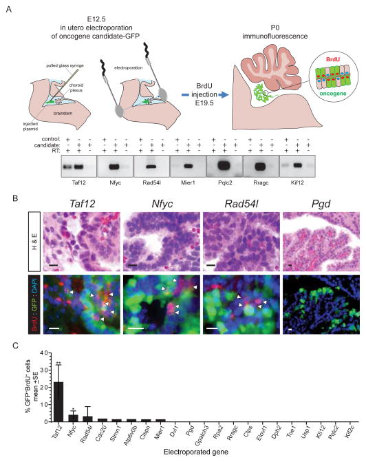
Figure 5. Functional in utero assessment of 1p31.3-ter/4qC6-qE2 candidate CPC oncogenes.
A. E12.5 choroid plexus was co-electroporated with plasmids encoding candidate and GFP to identify electroporated cells. Gels below show seven examples of 21 reverse-transcriptase (RT) PCR results of cells transfected with control plasmid or oncogene candidates with or without RT. B. Sections from each animal were analysed both for morphological change (top, H&E) and proliferation of electroporated cells (bottom, BrdU+/GFP+; scale bar=10μm). Only Taf12, Nfyc and Rad54l demonstrated dysplasia and aberrant proliferation relative to the other 18 candidates, Pgd shown as an example. C, Graph reporting the percentage of electroporated (GFP+) and proliferating choroid plexus epithelium cells. *P<0.05, **=P<0.005, Mann-Whitney.

