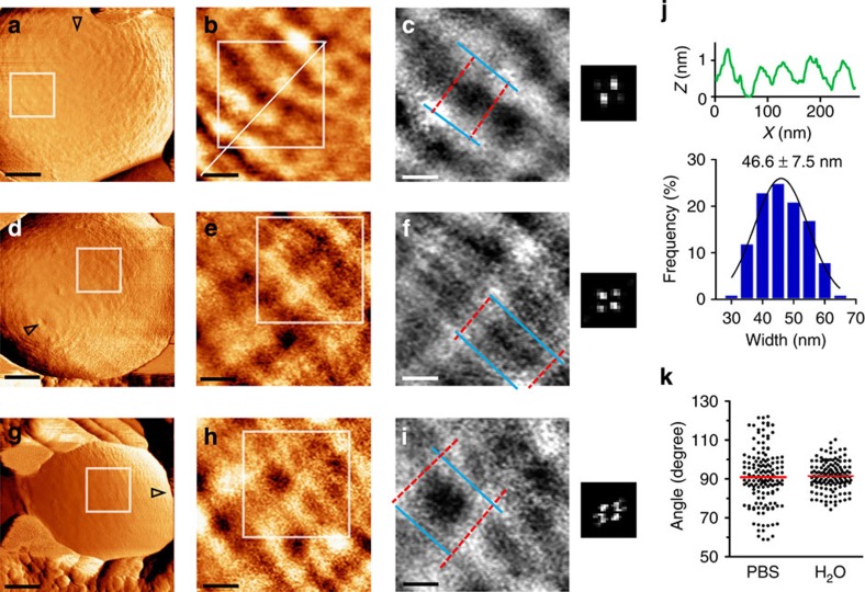
Figure 5. High turgor pressure stretches the peptidoglycan net.
(a,d,g) Low-magnification force-error images recorded in ultrapure water (scale bar, 200 nm; open triangles point towards the pole). (b,e,h) Height images of surface structure (scale bar, 50 nm; height, 2 nm). (c,f,i) High-magnification height images show larger holes in the PG net (scale bars, 30 nm; height, 1.5 nm). Corresponding 2D-FFT maps are at the right side of each image. (j) Representative height profile (upper) from a cross-section in b, and statistical analysis of band widths (histogram and an average±s.d.; n=15 bands per cell, from N=18 bacteria). (k) Angles plot shows similar average but narrower distribution range in water compared with PBS.

