Figure 1.
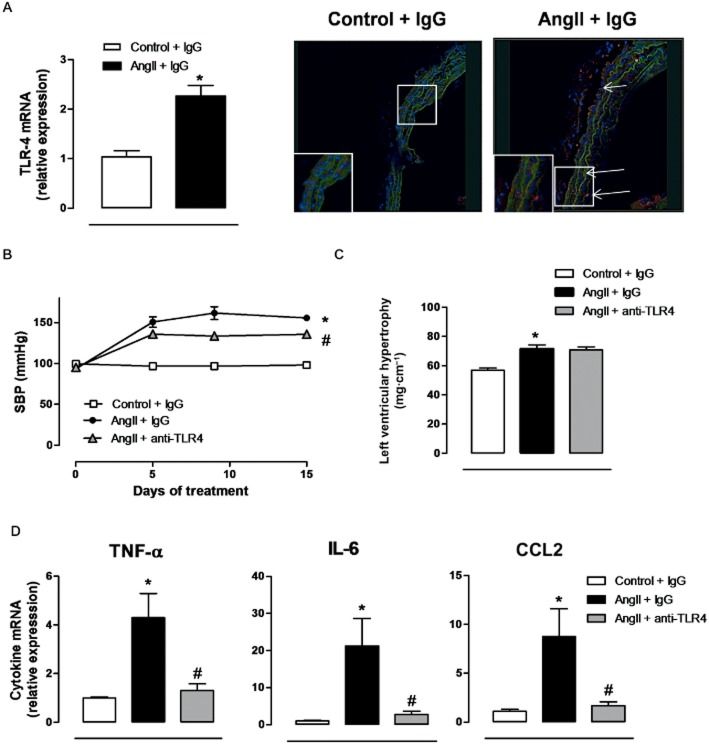
Increased TLR4 contributes to hypertension and inflammation in AngII-treated mice. (A) TLR4 mRNA levels and representative fluorescent confocal photomicrographs (×40 objective) of TLR4 immunolocalization in aortic segments from mice treated with a non-specific IgG and from mice treated with AngII plus a non-specific IgG. Image size: 375 × 375 μm. Positive immunostaining is indicated by arrows and the insets are magnified images of indicated areas. (B) SBP, (C) left ventricular hypertrophy and (D) levels of mRNA for TNF-α, IL-6 and CCL2 in mice treated with a non-specific IgG, with AngII plus a non-specific IgG or with AngII plus anti-TLR4 antibody. *P < 0.05 versus control + IgG, #P < 0.05 versus AngII + IgG. n = 6–16.

