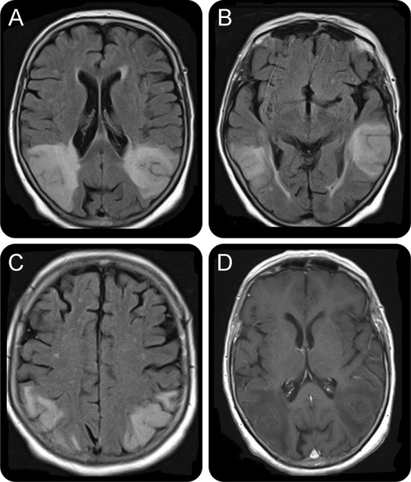
Figure. MRI brain 6 weeks post admission.

(A–C) Symmetrical high signal changes on fluid-attenuated inversion recovery sequences predominantly affecting the cortex, subcortical, and deep white matter in the posterior temporal and parieto-occipital regions. The areas of abnormal signal are wedge-shaped and extend to the ependymal surface of the lateral ventricles. There is gyral swelling and sulcal effacement. The limbic cortex is spared. (D) T1- weighted sequences plus contrast demonstrating lack of significant contrast enhancement. Of note, there was no diffusion restriction.
