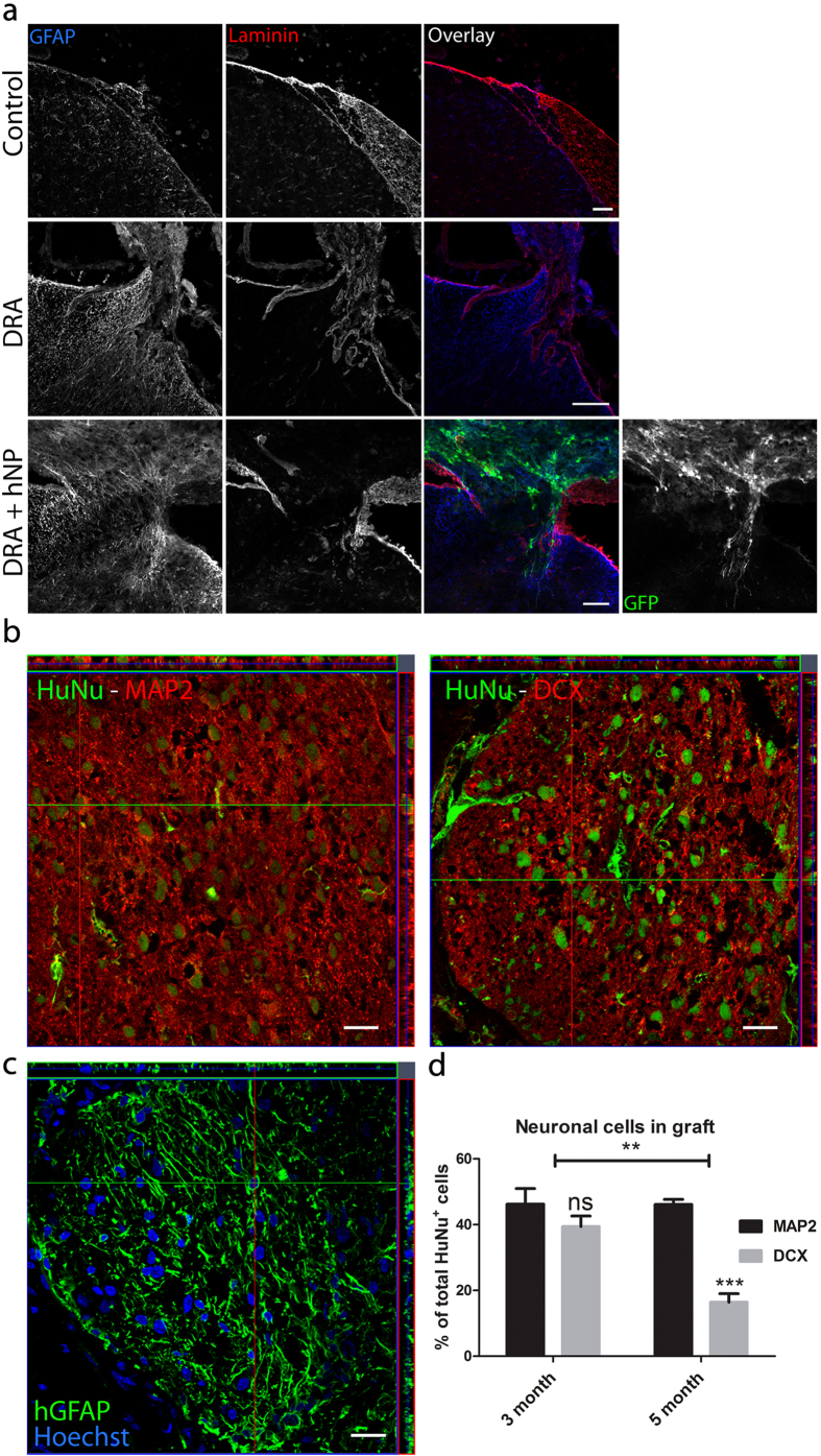
Figure 2. Engrafted hNP differentiated into neurons and glial cells at the site of dorsal root avulsion.
(a) The dorsal root entry zone was undisturbed by sham operations, whereas dorsal root avulsion (DRA) severely disrupted the spinal cord surface and led to the formation of a glial scar. Engrafted hNP were found in the vicinity of the injured dorsal horn associated with the surface of the spinal cord. Scale bar, 50 μm. (b+c) Engrafted hNP at the site of DRA gave rise to neurons characterized by the expression of MAP2 and DCX, and glial cells positive for human-specific GFAP. Scale bars, 25 μm. (d) three to five months after transplantation, hNP showed continuous neuronal differentiation indicated by decreasing expression of DCX and stable expression of MAP2. Y-axis shows percent of MAP2+ or DCX+ cells of the total number of HuNu+ cells per section. Data shown in d is in mean ± SEM of 3 animals per time point. Asterisks indicate level of statistical significance by two-way ANOVA with Sidak’s multiple comparison (**p < 0.01, ***p < 0.001).

