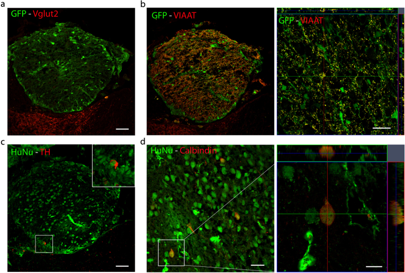
Figure 3. Engrafted human neurons differentiated primarily into inhibitory neurons.
(a) Engrafted hNP at the site of dorsal root avulsion (DRA) showed no immunoreactivity for Vglut2 . Scale bar, 50 μm. (b) Engrafted hNP at the site of DRA showed immunoreactivity for VIAAT localized to GFP+ boutons. Scale bars, 50 μm for the transplant overview, 20 μm for the orthogonal projection of a representative area in the transplant. (c) hNP at the site of DRA gave rise to a small number of TH+/HuNu+ cells. Inset shows a high power magnification of the region outlined by the white box. Scale bar, 50 μm. (d) Engrafted hNP at the site of DRA gave rise to calbindin+/HuNu+ interneurons. Scale bar, 25 μm for a representative area in the transplant, 10 μm for the orthogonal projection of the depicted calbindin+/HuNu+ cell.

