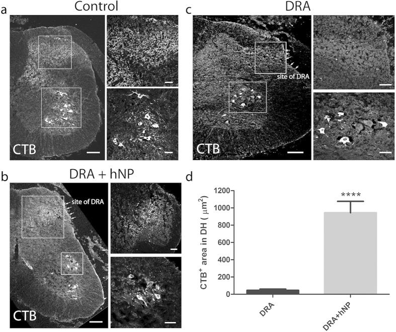
Figure 5. hNP transplantation led to the ingrowth of CTB labeled sensory fibers in the dorsal horn.
(a) CTB injected into the sciatic nerve of control animals was transganglionically transported to the ipsilateral dorsal horn (DH) and ventral motorneuron pools. (b) Animals that received hNP transplanted to the injury site of dorsal root avulsion (DRA) showed CTB labeling in the ipsilateral DH and ventral motorneurons. (c) In contrast, animals that underwent dorsal root avulsion (DRA) showed no transport of CTB into the DH but CTB was retrogradely transported to the ventral horn motorneuron pools. Scale bars, 100 μm for juxtaposed images depicting the ipsilateral side of the spinal cord, 50 μm for confocal pictures of ventral and DH areas. (d) Immunoreactivity for CTB in the ipsilateral DH along the site of DRA was exclusively found in animals receiving hNP transplants. Y-axis depicts CTB+ area in the ipsilateral DH per section. Data shown are in mean ± SEM of 3 animals per condition. Asterisks indicate level of statistical significance by student’s t-test (****p < 0.0001).

