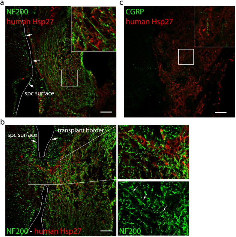
Figure 6. Engrafted hNP provided an environment for the ingrowth of sensory fibers.
(a) Engraftment sites of hNP showed a large number of parallel NF200+ fibers in close proximity to human cells. (b) NF200+ fibers in close proximity to human cells were able to grow into the dorsal horn. White arrows indicate NF200+ fibers passing from the hNP transplant into the spinal cord. (c) Only a very small number of CGRP+ fibers were found inside the transplant. Cells of human origin were identified by the cytoplasmic distribution of human specific Hsp27. Scale bars; 50 μm. Insets show a high power magnification of the regions outlined by the white box.

