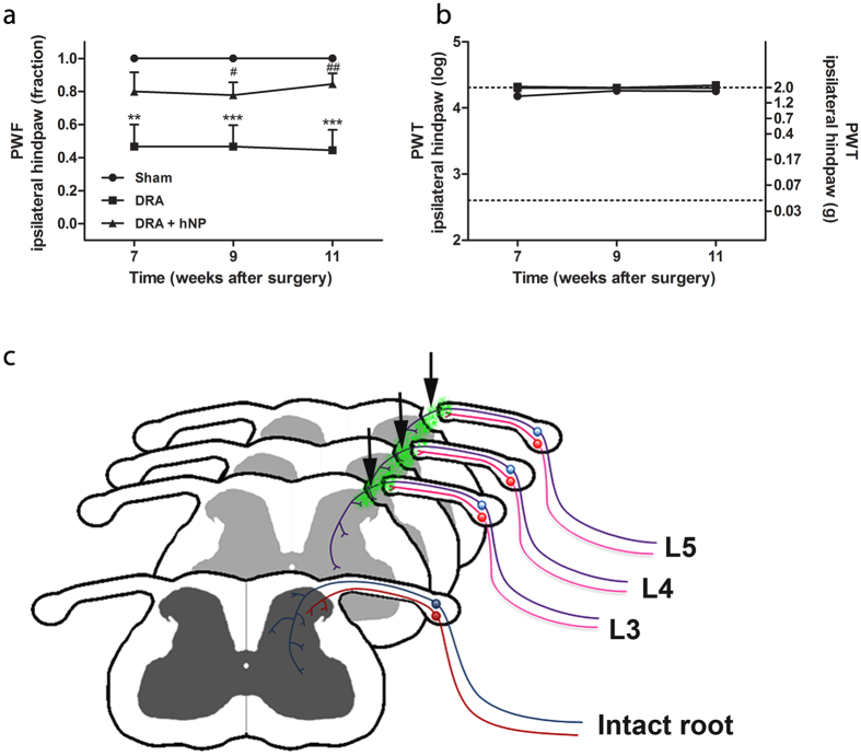
Figure 7. hNP transplantation partially rescued mechanical nociception in dorsal root avulsed mice.
(a) Dorsal root avulsion (DRA) strongly impaired the mechanical nociceptive function of the ipsilateral hind paw and transplantation of hNP significantly improved this function. (b) Neither DRA without treatment nor DRA with hNP transplantation showed mechanical allodynia. * indicates significant difference compared to sham, # indicates significant difference compared to DRA. PWF, paw withdrawal frequency; PWT, paw withdrawal threshold, data shown in a+b are in mean + SEM. Asterisks indicate level of statistical significance by one-way ANOVA with Tukey’s multiple comparison (#,*p < 0.05, ##,**p <0 .01, ***p < 0.001). (c) Graphical representation of the hNP graft localization and proposed regeneration of sensory fibers at the site of DRA. In the intact spinal cord myelinated (blue) and non-myelinated (red) sensory fibers project from the dorsal root ganglion via the dorsal root to the spinal cord dorsal horn (frontal image). After hNP (green) transplantation on the top of the avulsed and re-attached L3-L5 dorsal roots (arrows) myelinated fibers traverse the transplants and project to superficial and deep parts of the dorsal horn, whereas non-myelinated fibers grow into the transplant, but fail to enter the spinal cord.

