Figure 2.

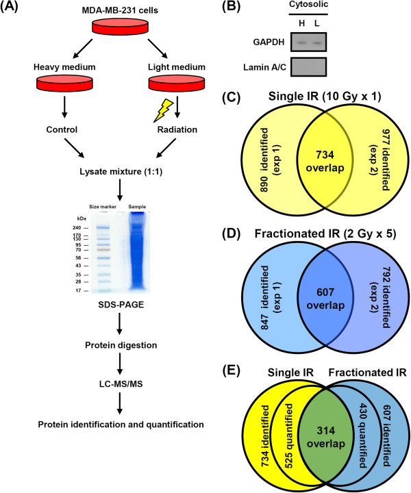
Venn diagram summary of identified proteins by SILAC-based quantitative proteomics. (A) Schematic workflow for profiling radiation-induced proteins via proteomic-based analysis. (B) The cytoplasmic lysates from SILAC-labeled cells were analyzed by western blotting to exclude contamination of nuclear extracts using GAPDH and Lamin A/C as cytosolic and nuclear control proteins, respectively. (C) 734 proteins were identified in the set of single dose experiments, and (D) 607 proteins were identified in the set of fractionated dose experiments. (E) Comparison of identified proteins from single or fractionated dose of irradiation.
