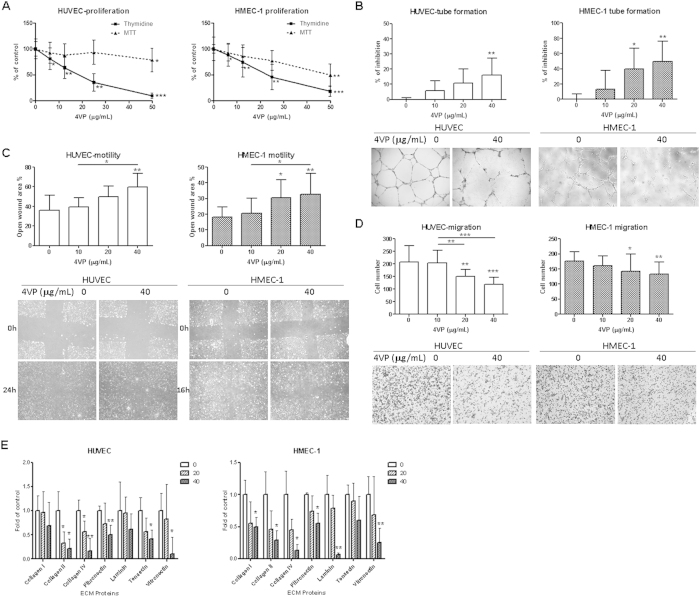
Figure 2. Effects of 4VP on cell proliferation, tube formation, migration, motility and cell adhesion to ECM proteins of HUVEC and HMEC-1.
(A) Cells were treated with increasing concentrations of 4VP for 48 hours, and cell proliferation was determined by the [methyl-3H] thymidine incorporation. Results are expressed as the mean % ratio of count per minute in treated and vehicle-treated control cells (mean ± SD of 4 independent experiments with 5 wells each). (B) Quantification of tube formation in HUVEC and HMEC-1 were shown. Representative photomicrographs showing the tube structures of HUVEC or HMEC-1 following 8 or 6 hours treatments, respectively, with vehicle (0 μg/mL) or indicated concentrations of 4VP. (C) Quantification of cell migration of HUVEC and HMEC-1 in Boyden chambers were shown. Representative photomicrographs showing the migrated and stained cells on the lower side of membranes. (D) Quantification of wound-induced cell motility in HUVEC and HMEC-1 were shown. Representative photomicrographs showing the cells migrated across the scratch wound in the presence or absence of 4VP after 16 or 24 hours incubation. (E) Effects of 4VP on cell adhesion to extracellular matrix proteins. Cells treated with 4VP (20 or 40 μg/mL) were added to a precoated plate containing different ECM proteins. The adhered cells were quantitated and results were shown. Results are expressed as the mean percentage of control (mean + SD of 3 independent experiments). Differences between the treated and vehicle-treated control groups were determined by one-way ANOVA with Tukey’s post-hoc test. Differences among treated groups were determined by one-way ANOVA with Tukey’s post-hoc test (B, C and D). *p < 0.05, **p < 0.01, ***p < 0.005 as compared among groups.

