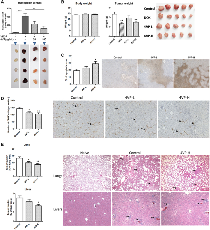
Figure 6. Inhibitory effects of 4VP on angiogenesis in mouse models.
(A) Upper: Hemoglobin content of Matrigel plugs from indicated groups (n = 10-11). Lower: The representative pictures of Matrigel plugs from indicated groups at day 7 after inoculation into mice. (B) The body and tumor weights of 4T1 tumor-bearing mice after Control (vehicle), 4VP-L (0.2 mg/kg) or 4VP-H (2 mg/kg) treatments. (C) Tumor apoptotic area and (D) endothelial cells in the tumor sections were assessed using TUNEL assay and CD31 immunohistochemical analysis, respectively. Representative photomicrographs in (D) showing the endothelial cells stained with anti-CD31 antibodies in brown. The paraffin-embedded sections of the lungs and livers were photographed and used to measure tumor area and total lung or liver area. The histograms showed the tumor burden in (E) lungs and (F) livers according to the tumor area as a percentage of whole lung or liver area per group. Representative H&E-stained sections of lungs and livers from different groups with arrows in (E) showing the tumor nodules (Naive group: no tumor inoculated and no treatment). Data are expressed as mean + SEM. Differences between the treated and vehicle-treated control groups were determined by one-way ANOVA with Tukey’s post-hoc test. *p < 0.05, **p < 0.01, ***p < 0.005 as compared among groups.

