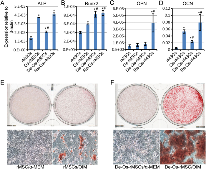
Figure 2. De-Os-MSCs showed osteogenic advantage in vitro.
(A) Total RNA were extracted from rMSCs, Os-rMSCs, De-Os-rMSCs and Re-Os-rMSCs. The relative expression levels of ALP, Runx2, OPN and OCN were checked by qRT-PCR. β-actin was used as an internal control. The data are expressed as mean ± SD (n = 3), *p < 0.05 compared to MSCs, #p < 0.05 compared to Os-MSCs. (B) Alizarin Red S staining of calcium deposits formed by MSCs. The untreated rMSCs and De-Os-rMSCs were cultured in α-MEM or osteogenic induction medium for 10 days, then the cells were fixed and stained with Alizarin Red S.

