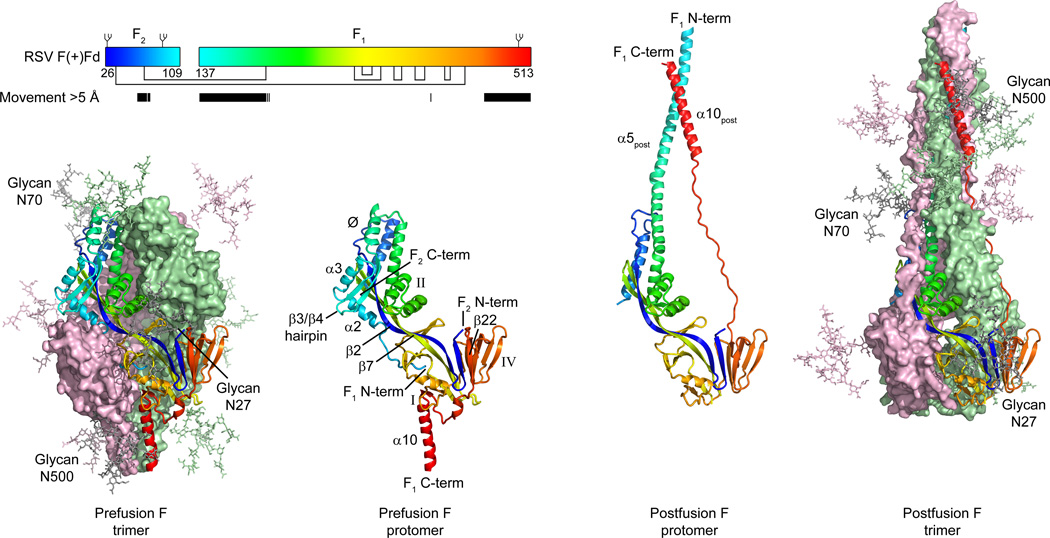
Figure 2. Structural rearrangement of RSV F.
To mediate virus-cell entry, the RSV F glycoprotein transitions from a metastable prefusion conformation to a stable postfusion conformation. Outer images display prefusion (left) and postfusion (right) trimeric structures, colored the same as in Fig. 1C. A complex glycan, shown as sticks, is modeled at each of the three N-linked glycosylation sites found in the mature protein. Inner images display a single RSV F protomer in ribbon representation, colored as a rainbow from blue to red, N-terminus of F2 to C-terminus of F1, respectively. Select secondary structure elements are labeled (correspondence with amino acid sequence in pre- and postfusion conformations is shown in fig. S3). Inset: schematic of the mature RSV F protein in the RSV F(+) Fd construct. The rainbow coloring of the boxes representing the F2 and F1 subunits matches that in the structures. Glycans are shown as branches on top of the boxes, and disulfide bonds are shown as black lines under the boxes. Amino acids that move more than 5 Å in the pre- and post-fusion conformations are indicated by black bars.

