
Figure 5.
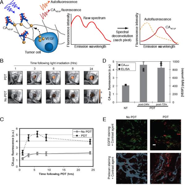
In vivo molecular imaging of cytokine secretion dynamics in response to PDT. Here, fluorescence hyperspectral imaging has been applied to acquire a secreted VEGF level time course in subcutaneous prostate cancer tumors (PC-3 human prostate cancer cells) following PDT. A. Schematic of concepts for molecular imaging of secreted VEGF levels in tumors using fluorescence hyperspectral imaging to extract contrast agent signal from autofluorescence background. B. Overlay of Avastin-Alexa Flour 680 conjugate (false colored gold; contrast agent for secreted VEGF, CAVEGF) fluorescence images (after spectral unmixing and calibration to a dye standard) and monochromatic reflectance images for a PDT-treated and an untreated control tumor. C. A time course of calibrated CAVEGF intensities for PDT-treated versus untreated control tumors. The full time course reveals a peak in VEGF secretion within 6 to 24 h post-PDT that returns to its pre-PDT baseline value after 3–7 days. D. Validation of VEGF imaging using tumor VEGF protein concentrations measured by ELISA on whole tumor lysates. The gray columns indicate mean calibrated CAVEGF intensity of untreated tumors (NT) and PDT-treated tumors at 24 h and 72 h following treatment; and, black diamonds indicate mean VEGF protein levels normalized by total protein concentration as determined by ELISA performed on whole tumor lysates. E. Localization of VEGF to cancer cells (green anti-EGFR stain) and heparin binding sites within the tumor ECM (blue, anti-perlecan stain) in ex vivo immunofluorescence images of cryosections from PDT-treated and untreated tumors. The mice were injected intravenously with CAVEGF (red) and treated with PDT 24 h prior to tumor harvesting and cryosectioning. Scale bar, 10 μm. The transient peak in secreted VEGF levels represents an opportune and critical time period for inhibiting VEGF activity. Adapted from Chang et al (2008).47