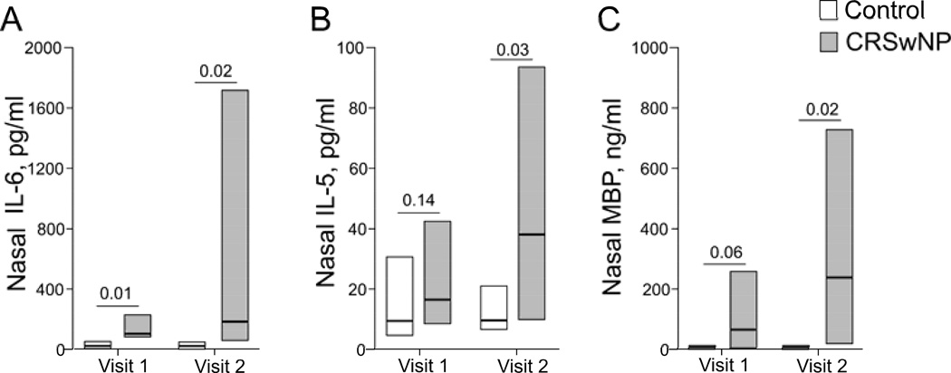
Figure 3.
Nasal levels of IL-6, IL-5 and MBP before (Visit 1) and during (Visit 2) acute exacerbation. Nasal secretions were collected before and during exacerbation and processed as described in the Methods section. Concentrations of inflammatory markers in the supernatants were analyzed by multiplex assay and radioimmunoassay. Data are presented as median (horizontal bars) and quartiles (boxes); n=10 for controls, n=9 for CRSwNP.

